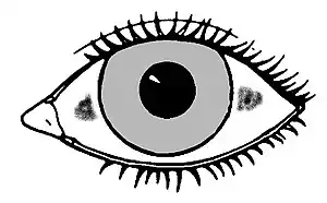
Bitot's spots
Bitot's spots are the buildup of keratin located superficially in the conjunctiva of human's eyes. They can be oval, triangular or irregular in shape. The spots are a sign of vitamin A deficiency and associated with drying of the cornea. In 1863, the French physician Pierre Bitot (1822–1888) first described these spots.[1] The spots may abate under replacement therapy.[2] In ancient Egypt, this was treated with animal liver, which is where vitamin A is stored.[3]
| Bitot's spots | |
|---|---|
| Other names | ICD10 = E50.1 |
 | |
| Typical location of Bitot's spots | |
| Specialty | Ophthalmology |
Causes
Major cause of Bitot's spot is vitamin A deficiency (VAD).[4] Rarely, pellegra due to deficiency of vitamin B3 (niacin) may also cause Bitot's spots.[5]
Treatment
VAD is commonly treated with oral vitamin A supplements.[6] Improvement of Bitot's spots is seen with high-dose vitamin A therapy.[7] Bitot's spots non-responsive to vitamin A therapy may be removed surgically.[8]
References
- Shukla, M; Behari, K (Jul 1979). "Congenital Bitot spots". Indian Journal of Ophthalmology. 27 (2): 63–4. PMID 541036.
- Ram, Jagat; Jinagal, Jitender (2018). "Bitot's Spots". New England Journal of Medicine. 379 (9): 869. doi:10.1056/NEJMicm1715354. PMID 30157394. S2CID 52126826.
- Numitor, Gerd (February 2012). Bitot's Spots. Flu Press. ISBN 978-620-0-57824-2.
- Gilbert, Clare (2013). "The eye signs of vitamin A deficiency". Community Eye Health. 26 (84): 66–67. ISSN 0953-6833. PMC 3936686. PMID 24782581.
- Levine, Robert A.; Rabb, Maurice F. (1 November 1971). "Bitot's Spot Overlying a Pinguecula". Archives of Ophthalmology. 86 (5): 525–528. doi:10.1001/archopht.1971.01000010527007. PMID 5315641.
- "Vitamin A Deficiency Treatment & Management: Medical Care, Consultations, Diet". 9 November 2019.
- "Management of Bitot's Spots". American Academy of Ophthalmology. 1 December 2016.
- Themes, U. F. O. (11 September 2016). "Bitot's Spots". Ento Key.
External links