Bregma
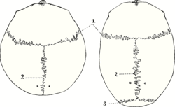
The bregma is the anatomical point on the skull at which the coronal suture is intersected perpendicularly by the sagittal suture.
| Bregma | |
|---|---|
 | |
| Details | |
| Precursor | anterior fontanelle |
| System | skeletal system |
| Identifiers | |
| Latin | bregma |
| TA98 | A02.1.00.016 |
| TA2 | 418 |
| FMA | 264776 |
| Anatomical terminology | |
Structure
The bregma is located at the intersection of the coronal suture and the sagittal suture on the superior middle portion of the calvaria.[1] It is the point where the frontal bone and the two parietal bones meet.[1]
Development
The bregma is known as the anterior fontanelle during infancy. The anterior fontanelle is membranous and closes in the first 18-36 months of life.[2]
Clinical significance
Cleidocranial dysostosis
In the birth defect cleidocranial dysostosis, the anterior fontanelle never closes to form the bregma.
Surgical landmark
The bregma is often used as a reference point for stereotactic surgery of the brain.[3][4] It may be identified by blunt scraping of the surface of the skull and washing to make the meeting point of the sutures clearer.[3]
Neonatal examination
Examination of an infant includes palpating the anterior fontanelle.[5] It should be flat, soft, and less than 3.5cm across.[5] A sunken fontanelle indicates dehydration, whereas a very tense or bulging anterior fontanelle indicates raised intracranial pressure.
Height assessment
Cranial height is defined as the distance between the bregma and the midpoint of the foramen magnum (the basion).[6] This is strongly linked to more general growth.[6] This can be used to assess the general health of a deceased person as part of an archaeological excavation, giving information on the health of a population.[6]
Etymology
The word "bregma" comes from the Ancient Greek βρέγμα (brégma), meaning the bone directly above the brain.[7]
References
![]() This article incorporates text in the public domain from page 135 of the 20th edition of Gray's Anatomy (1918)
This article incorporates text in the public domain from page 135 of the 20th edition of Gray's Anatomy (1918)
- "Skull, Scalp, and Meninges Overview". Imaging in Neurology, Part 1. AMIRSYS. 2016. pp. 288–291. doi:10.1016/B978-0-323-44781-2.50232-1. ISBN 978-0-323-44781-2.
- Gilroy, Anne M.; MacPherson, Brian R.; Wikenheiser, Jamie C.; Schuenke, Michael; Schulte, Erik; Schumacher, Udo (2020). Atlas of Anatomy. Anne M. Gilroy, Brian R. MacPherson, Jamie C. Wikenheiser, Markus M. Voll, Karl Wesker, Michael Based on: Schünke (4th ed.). New York: Thieme Medical Publishers. ISBN 978-1-68420-203-4. OCLC 1134458436.
{{cite book}}: CS1 maint: date and year (link) - Carvey, Paul M.; Maag, Terrence J.; Lin, Donghui (1994). "13 - Injection of Biologically Active Substances into the Brain". Methods in Neurosciences. Vol. 21. Elsevier. pp. 214–234. doi:10.1016/B978-0-12-185291-7.50019-9. ISBN 978-0-12-185291-7. ISSN 1043-9471.
- Harley, Carolyn W.; Shakhawat, Amin M. D.; Quinlan, Meghan A. L.; Carew, Samantha J.; Walling, Sue G.; Yuan, Qi; Martin, Gerard M. (2018). "Chapter 19 - Using Molecular Biology to Address Locus Coeruleus Modulation of Hippocampal Plasticity and Learning: Progress and Pitfalls". Handbook of Behavioral Neuroscience. Vol. 28. Publisher. pp. 349–364. doi:10.1016/B978-0-12-812028-6.00019-7. ISBN 978-0-12-812028-6. ISSN 1569-7339.
- Carreiro, Jane E. (2009-01-01). "8 - Labor, delivery and birth". An Osteopathic Approach to Children (2nd ed.). Churchill Livingstone. pp. 131–145. doi:10.1016/b978-0-443-06738-9.00008-3. ISBN 978-0-443-06738-9.
{{cite book}}: CS1 maint: date and year (link) - Nikita, Efthymia (2017-01-01). "6 - Growth Patterns". Osteoarchaeology - A Guide to the Macroscopic Study of Human Skeletal Remains. Academic Press. pp. 243–267. ISBN 978-0-12-804021-8.
{{cite book}}: CS1 maint: date and year (link) - Liddell & Scott, Greek-English Lexicon
Additional images
 The bregma, human skull.
External links
- lesson1 at The Anatomy Lesson by Wesley Norman (Georgetown University)