Clivus (anatomy)
The clivus (/ˈklaɪvəs/,[1] Latin for "slope"), or Blumenbach clivus, is a bony[2] part of the cranium at the base of the skull. It is a shallow depression behind the dorsum sellae of the sphenoid bone. It slopes gradually to the anterior part of the basilar occipital bone at its junction with the sphenoid bone. It extends to the foramen magnum. It is related to the pons and the abducens nerve (CN VI).
| Clivus | |
|---|---|
 Superior view of the clivus | |
| Details | |
| Identifiers | |
| Latin | Clivus |
| TA98 | A02.1.00.051 A02.1.04.006 |
| TA2 | 454 |
| FMA | 54376 |
| Anatomical terms of bone | |
Structure
The clivus is a shallow depression behind the dorsum sellae of the sphenoid bone.[3] It slopes gradually to the anterior part of the basilar occipital bone at its junction with the sphenoid bone. It extends to the foramen magnum.[3] On axial planes, it sits just posterior to the sphenoid sinuses. It is medial to the foramen lacerum (the internal carotid artery reaches the middle cranial fossa above the foramen lacerum), proximal to its anastomosis with the Circle of Willis. It is anterior to the basilar artery.
The pons sits on the clivus.[3] The abducens nerve (CN VI) also tracks along the clivus during its course.[3]
Variations
During embryonic development, the clivus is formed by the fusion of the basiocciput and basisphenoid or also known as the sphenooccipital synchondrosis. When the fusion occurs improperly, it would give rise to gaps that are considered anatomical variations. Variations of the clivus include fossa navicularis magna, craniopharyngeal canal, canalis basilaris medianus, and transverse basilar fissure (Saucer's fissure).[4] Ossification of the apical ligament of dens may also occur, resulting in a variant bony tubercle at the inferior end of the clivus.[4] Condylus tertius and arcus praebasiocipitalis are the other two variations that can be found at the lower end of the clivus, although their etiology may be different from the other variations. Ecchordosis physaliphora, a congenital benign lesion derived from the notochord, might be present in the dorsal part of the clivus.[5] This lesion is harmless is considered an anatomical variant.
Clinical importance
The abducens nerve (CN VI) tracks along the clivus during its course.[3] Increased intracranial pressure can trap the nerve at this point and cause signs of palsy.
The clivus is also the site for chordoma, a rare type of cancer.
Surgery
Surgery for lesions involving the clivus and surrounding structures have traditionally been approached via extended subfrontal transbasal, anterior transfacial, lateral transtemporal, far-lateral approaches, and staged approaches.[6] These approaches are limited in that they often require extensive bone removal and brain retraction while placing critical neurovascular structures between the surgeon and the site of pathology. It has been proposed that these limitations are mitigated by significant advancements in the use of endoscopic endonasal surgery. Contemporary surgical approaches involving extended endoscopic endonasal approaches to the clivus have been increasingly described by several groups, and have been shown to be a safe and effective strategy for the surgical management of a variety of benign and malignant lesions.[6]
Relation of the clivus and dens
The clivus is an important landmark for checking for anatomical atlanto-occipital alignment. Wheen viewed on a lateral C-spine radiograph, the clivus forms a line which, if extended, is known as Wackenheim's clivus line. Wackenheim's clivus line should pass through the dens of the axis or be tangential to it.[7]
History
"Clivus" is also used as an abbreviated term for the clivus ocularis, which is the sloping inner wall of the retina as it dips into the foveola in the macula of the eye. To disambiguate, the clivus is sometimes referred to as the Blumenbach clivus. This is named after Johann Friedrich Blumenbach.
Additional images
 Clivus
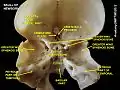 Clivus
See also
References
![]() This article incorporates text in the public domain from page 148 of the 20th edition of Gray's Anatomy (1918)
This article incorporates text in the public domain from page 148 of the 20th edition of Gray's Anatomy (1918)
- "Definition of clivus | Dictionary.com". www.dictionary.com. Retrieved 2022-01-05.
- Drake, Richard L. Gray's Anatomy for Students 3rd Ed. p. 868.
- Maira, Giulio; Doglietto, Francesco; Pallini, Roberto (2012). "41 - Surgical Management of Lesions of the Clivus". Schmidek and Sweet Operative Neurosurgical Techniques (6th ed.). Saunders. pp. 486–500. doi:10.1016/B978-1-4160-6839-6.10041-3. ISBN 978-1-4160-6839-6.
- Hofmann, E.; Prescher, A. (2012-06-01). "The Clivus". Clinical Neuroradiology. 22 (2): 123–139. doi:10.1007/s00062-011-0083-4. ISSN 1869-1447.
- Lagman, Carlito; Varshneya, Kunal; Sarmiento, J. Manuel; Turtz, Alan R.; Chitale, Rohan V. (2016-03-30). "Proposed Diagnostic Criteria, Classification Schema, and Review of Literature of Notochord-Derived Ecchordosis Physaliphora". Cureus. 8 (3). doi:10.7759/cureus.547. ISSN 2168-8184.
- Little, Ryan E.; Taylor, Robert J.; Miller, Justin D.; Ambrose, Emily C.; Germanwala, Anand V.; Sasaki-Adams, Deanna M.; Ewend, Matthew G.; Zanation, Adam M. (August 2014). "Endoscopic endonasal transclival approaches: case series and outcomes for different clival regions". Journal of Neurological Surgery. Part B, Skull Base. 75 (4): 247–254. doi:10.1055/s-0034-1371522. ISSN 2193-6331. PMC 4108492. PMID 25093148.
- McKenna DA, Roche CJ, Lee KW, Torreggiani WC, Duddalwar VA. Atlanto-occipital dislocation: case report and discussion. Can J Emerg Med 2006; 8(1):50-3. Available at: link Archived 2007-09-27 at the Wayback Machine and link Archived 2007-09-27 at the Wayback Machine. Accessed on: December 7, 2006.
External links
- Anatomy photo:22:os-0913 at the SUNY Downstate Medical Center - "Osteology of the Skull: Internal Surface of Skull"
- Diagram at uwo.ca