Parasitic twin
A parasitic twin, also known as an asymmetrical or unequal conjoined twin, is the result of the processes that also produce vanishing twins and conjoined twins, and may represent a continuum between the two. Parasitic twins occur when a twin embryo begins developing in utero, but the pair does not fully separate, and one embryo maintains dominant development at the expense of its twin. Unlike conjoined twins, one ceases development during gestation and is vestigial to a mostly fully formed, otherwise healthy individual twin. The undeveloped twin is defined as parasitic, rather than conjoined, because it is incompletely formed or wholly dependent on the body functions of the complete fetus.[1] The independent twin is called the autosite.
| Parasitic twin | |
|---|---|
 | |
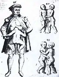
| Illustration of a man with a parasitic twin, alongside illustrations of two configurations of conjoined twins | |
| Specialty | Maternal–fetal medicine, neonatology |
Variants
- Conjoined parasitic twins joined at the head are described as craniopagus or cephalopagus, and occipitalis if joined in the occipital region or parietalis if joined in the parietal region.
- Craniopagus parasiticus is a general term for a parasitic head attached to the head of a more fully developed fetus or infant.[2]
- Fetus in fetu sometimes is interpreted as a special case of parasitic twin, but may be a distinct entity.
- The twin reversed arterial perfusion, or T.R.A.P. sequence, results in an 'acardiac twin', a parasitic twin that fails to develop a head, arms and a heart. The parasitic twin, little more than a torso with or without legs, receives its blood supply from the host twin by means of an umbilical cord-like structure, much like a fetus in fetu, except the acardiac twin is outside the autosite's body. The blood received by the parasitic twin has already been used by the normal fetus, and as such is already de-oxygenated, leaving little developmental nutrients for the acardiac twin. Because it is pumping blood for both itself and its acardiac twin, this causes extreme stress on the normal fetus' heart. Many T.R.A.P. pregnancies result in heart failure for the healthy twin. This twinning condition usually occurs very early in pregnancy.[3] A rare variant of the acardiac fetus is the acardius acormus where the head is well developed but the heart and the rest of the body are rudimentary. While it is thought that the classical T.R.A.P./Acardius sequence is due to a retrograde flow from the umbilical arteries of the pump twin to the iliac arteries of the acardiac twin resulting in preferential caudal perfusion, acardius acormus is thought to be a result of an early embryopathy.[4]
Images of parasitic twins
Human
 40-year-old man seen in Paris, 1530
 30-year-old Neapolitan man, seen in 1742
 Indian man, 1787
.jpg.webp) Agan, a Chinese man, 1833
Agan, a Chinese man, 1833 9-year-old Gustav Evrard of Paris, 1839
 Blanche Dumas of France, born 1860
 Joao Baptista dos Santos and Louise L. ("la femme de quatre jambes"), 1893
Animals
 Chicken with 2 extra lower limbs
 Pigeon with 2 extra lower limbs
 Cat with lower body duplication
 Sheep with 2 extra front limbs
See also
- Dipygus
- Frank Lentini
- Lakshmi Tatma
- Lazarus and Joannes Baptista Colloredo
- Rudy Santos
- Vestigial twin
References
- "Parasitic Twins | The Embryo Project Encyclopedia". embryo.asu.edu. Retrieved 2022-02-18.
- Aquino DB, Timmons C, Burns D, Lowichik A (1997). "Craniopagus parasiticus: a case illustrating its relationship to craniopagus conjoined twinning". Pediatric Pathology and Laboratory Medicine. 17 (6): 939–44. doi:10.1080/107710497174381. PMID 9353833.
- "Acardiac Twin or TRAP Sequence". University of California, San Francisco. 2007-04-26. Archived from the original on 2012-07-08. Retrieved 2007-05-30.
- Abi Nader Khalil, Whitten Sara Melissa, Filippi Elisa, Scott Rose-Mary, Jauniaux Eric. Dichorionic triamniotic triplet pregnancy complicated by acardius acormus. Fetal Diagnosis and Therapy 2009;26(1):45-9.
Further reading
- Lebel, Robert Roger; Carlos Mock; Jeannette Israel; William Senica (June 1994). "Twin reversed arterial perfusion syndrome, acephalic presenting at full term". TheFetus.net. Retrieved 18 February 2013.
This article is issued from Wikipedia. The text is licensed under Creative Commons - Attribution - Sharealike. Additional terms may apply for the media files.