Subungual exostosis
Subungual exostoses are a type of non-cancerous bone tumor of the chondrogenic type, and consists of bone and cartilage.[2] It usually projects from the upper surface of the big toe underlying the nailbed, giving rise to a painful swelling that destroys the nail.[3] Subsequent ulceration and infection may occur.[3]
| Subungual exostosis | |
|---|---|
| Other names | Dupuytren subungual exostosis[1] |
 | |
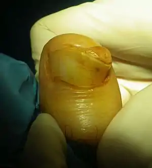
| Subungual exostosis (1/3), in a boy age 15 years | |
| Specialty | Orthopedics |
There is an association with trauma and infection.[4] Diagnosis involves medical imaging to exclude other similar conditions, particularly osteochondroma.[5] X-ray appearance may reveal a bony protuberance attached to the top or side surface of a toe bone.[6]
Treatment is by surgical excision and is effective.[6]
More than half are under the age of 18 years and males are affected equally to females.[3] Combined with bizarre parosteal osteochondromatous proliferation, they comprise <5% of cartilage tumors.[4]
Signs and symptoms
They tend to be painful due to the pressure applied to the nail bed and plate. They can involve destruction of the nail bed.[7] These lesions are not true osteochondromas, rather it is a reactive cartilage metaplasia. The reason it occurs on the dorsal aspect is because the periosteum is loose dorsally but very tightly adherent volarly.[8]
Diagnosis
Diagnosis involves medical imaging.[5]
Differential diagnosis includes mainly bizarre parosteal osteochondromatous proliferation (BPOP), which is more irregular and tends to involve the middle of the finger or toe rather than the end near the nail.[6] They are distinct from subungual osteochondroma.[9]
Treatment
Treatment is by surgical excision and is effective.[6]
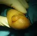
 Subungual exostosis (2/3)
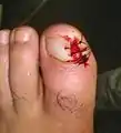
 Subungual exostosis (3/3), after excision
Epidemiology
It tends to occur in children and adolescents. Combined with BPOP, they account for less than 5% of cartilage tumors.[4]
References
- "Dupuytren subungual exostosis | Genetic and Rare Diseases Information Center (GARD) – an NCATS Program". rarediseases.info.nih.gov. Retrieved 11 October 2017.
- WHO Classification of Tumours Editorial Board, ed. (2020). "Bone tumors". Soft Tissue and Bone Tumours: WHO Classification of Tumours. Vol. 3 (5th ed.). Lyon (France): International Agency for Research on Cancer. p. 338. ISBN 978-92-832-4503-2.
- WHO Classification of Tumours Editorial Board, ed. (2020). "Subungual exostosis". Soft Tissue and Bone Tumours: WHO Classification of Tumours. Vol. 3 (5th ed.). Lyon (France): International Agency for Research on Cancer. pp. 345–347. ISBN 978-92-832-4503-2.
- Engel, Hannes; Herget, Georg W.; Füllgraf, Hannah; Sutter, Reto; Benndorf, Matthias; Bamberg, Fabian; Jungmann, Pia M. (March 2021). "Chondrogenic Bone Tumors: The Importance of Imaging Characteristics". RöFo: Fortschritte auf dem Gebiete der Röntgenstrahlen und der Nuklearmedizin. 193 (3): 262–275. doi:10.1055/a-1288-1209. ISSN 1438-9010. PMID 33152784.
- DaCambra, Mark P.; Gupta, Sumit K.; Ferri-de-Barros, Fabio (April 2014). "Subungual Exostosis of the Toes: A Systematic Review". Clinical Orthopaedics and Related Research. 472 (4): 1251–1259. doi:10.1007/s11999-013-3345-4. ISSN 0009-921X. PMC 3940761. PMID 24146360.
- Bocklage, Therese J.; Quinn, Robert; Verschraegen, Claire; Schmit, Berndt (2014). "16. Cartilaginous tumours of bones and joints". Bone and Soft Tissue Tumors: A Multidisciplinary Review with Case Presentations. London: JP Medical Ltd. p. 379. ISBN 978-1-907816-22-2.
- Suga H, Mukouda M (2005). "Subungual exostosis: a review of 16 cases focusing on postoperative deformity of the nail". Annals of Plastic Surgery. 55 (3): 272–5. doi:10.1097/01.sap.0000174356.70048.b8. PMID 16106166. S2CID 5813472.
- Murphey MD, Choi JJ, Kransdorf MJ, et al: Imaging of osteochondroma: variants and complications with radiologic-pathologic correlation. Radiographics 20:1407-1434, 2000
- Lee SK, Jung MS, Lee YH, Gong HS, Kim JK, Baek GH (2007). "Two distinctive subungual pathologies: subungual exostosis and subungual osteochondroma". Foot & Ankle International. 28 (5): 595–601. doi:10.3113/FAI.2007.0595. PMID 17559767. S2CID 41309941.
External links