Thyrohyoid muscle
The thyrohyoid muscle is a small skeletal muscle on the neck. It originates from the lamina of the thyroid cartilage, and inserts into the greater cornu of the hyoid bone. It is supplied by the hypoglossal nerve, and a branch of the ventral rami of the cervical plexus, spinal nerve C1, which travels with the hypoglossal nerve. The thyrohyoid muscle depresses the hyoid bone and elevates the larynx. By controlling the position and shape of the larynx, it aids in making sound.
| Thyrohyoid muscle | |
|---|---|
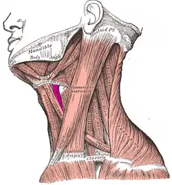 Muscles of the neck. Lateral view. (Thyrohyoideus labeled center-left.) | |
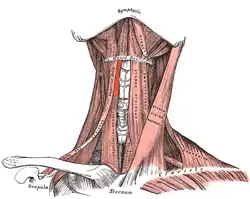 Muscles of the neck. Anterior view. (Thyrohyoideus visible center-left.) | |
| Details | |
| Origin | thyroid cartilage of the larynx |
| Insertion | hyoid bone |
| Artery | superior thyroid artery |
| Nerve | hypoglossal nerve, first cervical nerve (C1) via hypoglossal nerve |
| Actions | elevates thyroid and depresses the hyoid bone |
| Identifiers | |
| Latin | Musculus thyrohyoideus |
| TA98 | A04.2.04.007 |
| TA2 | 2174 |
| FMA | 13344 |
| Anatomical terms of muscle | |
Structure
The thyrohyoid muscle is a quadrilateral muscle in shape. It appears like an upward continuation of the sternothyroid muscle. It belongs to the infrahyoid muscles group. It lies in the carotid triangle.[1]
It arises from the oblique line on the lamina of the thyroid cartilage.[2] It is inserted into the lower border of the greater cornu of the hyoid bone.[2]
Nerve supply
The thyrohyoid muscle is supplied by the hypoglossal nerve (XII).[3] It is the only infrahyoid muscle that is not supplied by the ansa cervicalis.[4] It is also supplied by the thyrohyoid branch of cervical spinal nerve 1 (C1).[5] This is via the cervical plexus.[6] This nerve branches from the first cervical nerve as it joins the hypoglossal nerve for a short distance.[5]
Function
The thyrohyoid muscle depresses the hyoid bone and elevates the larynx and the thyroid cartilage, drawing them together.[2] By controlling the position and shape of the larynx, it aids in making sound.[7]
Other animals
The thyrohyoid muscle is found in many other animals, including horses.[2]
Additional images
 Hyoid bone. Anterior surface. Enlarged.
 The veins of the thyroid gland.
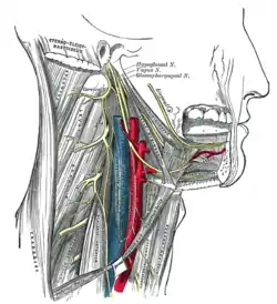 Hypoglossal nerve, cervical plexus, and their branches.
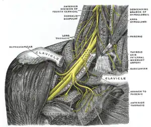 The right brachial plexus with its short branches, viewed from in front.
 Side view of the larynx, showing muscular attachments.
 Thyrohyoid muscle
See also
References
![]() This article incorporates text in the public domain from page 394 of the 20th edition of Gray's Anatomy (1918)
This article incorporates text in the public domain from page 394 of the 20th edition of Gray's Anatomy (1918)
- Luna, Mario A.; Pfaltz, Madeleine (2009). "11 - Cysts of the Neck, Unknown Primary Tumor, and Neck Dissection". Diagnostic Surgical Pathology of the Head and Neck (2nd ed.). Philadelphia: Saunders. pp. 839–881. doi:10.1016/B978-1-4160-2589-4.00011-5. ISBN 978-1-4377-1951-2. OCLC 460904310.
- Parente, Eric J.; Franklin, Samantha H.; Derksen, Frederik J.; Weishaupt, Michael A.; Chalmers, Heather J.; Tessier, Caroline (2012). "42 - Diagnostic Techniques in Equine Upper Respiratory Tract Disease". Equine Surgery (4th ed.). St. Louis: Saunders. pp. 536–557. doi:10.1016/B978-1-4377-0867-7.00042-9. ISBN 978-1-4377-0868-4. OCLC 761714103.
- Thomas, P. K.; Mathias, Christopher J. (2005). "52 - Diseases of the Ninth, Tenth, Eleventh, and Twelfth Cranial Nerves". Peripheral neuropathy (4th ed.). Philadelphia: Saunders. pp. 1273–1293. doi:10.1016/B978-0-7216-9491-7.50055-7. ISBN 978-1-4377-1347-3. OCLC 324998243.
- Netter, Frank H. (2018). Atlas of Human Anatomy. Elsevier. pp. Table 2.9. ISBN 9780323393218.
- Brandmeir, Nicholas (2015). "32 - Nerve Injuries of the Neck". Nerves and nerve injuries: Pain, treatment, injury, disease and future directions. Vol. 2. London, UK: Academic Press. pp. 493–504. doi:10.1016/B978-0-12-802653-3.00081-6. ISBN 0-12-802653-7. OCLC 908203065.
- "Thyrohyoid muscle".
- Hage, Steffen R. (2010). "8.3 - Neuronal networks involved in the generation of vocalization". Handbook of Behavioral Neuroscience. Stefan Brudzynski. London: Academic Press. pp. 339–349. doi:10.1016/B978-0-12-374593-4.00032-2. ISBN 978-0-12-374593-4. ISSN 1569-7339. OCLC 528610774.
External links
- Anatomy photo:25:03-0106 at the SUNY Downstate Medical Center
- Anatomy photo:25:10-0105 at the SUNY Downstate Medical Center
- "Anatomy diagram: 25420.000-1". Roche Lexicon - illustrated navigator. Elsevier. Archived from the original on 2015-02-26.
- PTCentral