Pharyngobasilar fascia
As it descends it diminishes in thickness, and is gradually lost.
| Pharyngobasilar fascia | |
|---|---|
| Details | |
| Identifiers | |
| Latin | fascia pharyngobasilaris |
| TA98 | A05.3.01.027 |
| TA2 | 2858 |
| FMA | 55074 |
| Anatomical terminology | |
It is strengthened posteriorly by a strong fibrous band, which is attached above to the pharyngeal spine on the under surface of the basilar portion of the occipital bone, and passes downward, forming a median raphé, which gives attachment to the Constrictores pharyngis.
Additional images
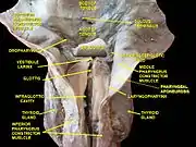 Larynx, pharynx and tongue.Deep dissection, posterior view.
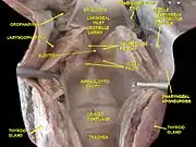 Larynx, pharynx and tongue.Deep dissection, posterior view.
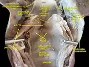 Larynx, pharynx and tongue.Deep dissection, Posterior view.
References
![]() This article incorporates text in the public domain from page 1143 of the 20th edition of Gray's Anatomy (1918)
This article incorporates text in the public domain from page 1143 of the 20th edition of Gray's Anatomy (1918)
External links
- "Pharyngobasilar fascia". Medcyclopaedia. GE. Archived from the original on 2012-02-05.
- http://ect.downstate.edu/courseware/haonline/labs/l31/100101.htm
- http://www.instantanatomy.net/headneck/areas/phpharyngobasilarfascia.html
This article is issued from Wikipedia. The text is licensed under Creative Commons - Attribution - Sharealike. Additional terms may apply for the media files.