Retropharyngeal space
The retropharyngeal space (abbreviated as "RPS"[1][2]) is a potential space[2] and deep compartment of the head and neck[1] situated posterior to the pharynx.[3] The RPS is bounded anteriorly by the buccopharyngeal fascia, posteriorly by the prevertebral fascia, and laterally by the carotid sheath. It spans from the base of the skull superiorly to the mediastinum inferiorly.[1] It contains the retropharyngeal lymph nodes.[2]
| Retropharyngeal space | |
|---|---|
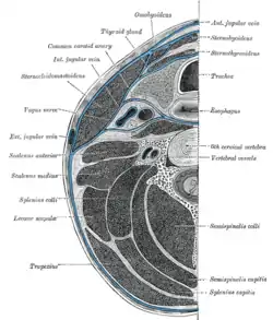 Section of the neck at about the level of the sixth cervical vertebra. Showing the arrangement of the deep cervical fascia | |
 | |
| Details | |
| Identifiers | |
| Latin | Spatium retropharyngeum |
| TA98 | A05.3.01.118 |
| TA2 | 2884 |
| FMA | 84965 |
| Anatomical terminology | |
Sources consider the retropharyngeal space to be in principle subdivided into the so-called "true retropharyngeal space"[1][4] or "retropharyngeal space proper" (part of the RSP situated anterior to the alar fascia),[4][2] and the danger space (part of the RSP situated posterior to the alar fascia).[1][2][4] The danger space is sometimes also lumped together with the true RPS and the whole referred to as the RPS because the alar fascia is an ineffective barrier.[2] Infections from the head and neck can spread down through the danger space into the posterior mediastinum.[2]
Anatomy
Superiorly, the retropharingeal space terminates at the base of the skull (more specifically, at the clivus[2]).[1][4] Inferiorly, the true RPS terminates at a variable level along the upper thoracic spine with the fusion of alar fascia and visceral fascia;[1] sources either give the inferior termination of the true RPS as occurring at approximately the vertebral level of T4[2] or at a variable level anywhere between the T1-T6.[1][4] The danger space component of the RPS meanwhile extends further inferior-ward, entering the posterior mediastinum to reach the level of the diaphragm.[1]
Contents
The retropharnygeal space contains the retropharyngeal lymph nodes.[2] The suprahyoid portion of the RPS contains the lymph nodes as well as adipose tissue, while the infrahyoid portion contains adipose tissue only.[1]
A midline raphe is sometimes present in the RPS, subdividing it into a left half and a right half.[2]
Anatomical relations
Positions of adjacent anatomical structures in relation to the retropharyngeal space are as follows:
- Superior: Base of the skull[1][5]
- Inferior: Posterior mediastinum[5]
- Lateral: Carotid sheath[1]
- Anterior: Buccopharyngeal fascia[1]
- Posterior: Prevertebral fascia (alar fascia true RPS)[1]
Clinical significance
A midline raphe may be present in this the RPS,[2] making some infections appear unilateral. However without treatment infections can easily spread from one space to the adjacent space.
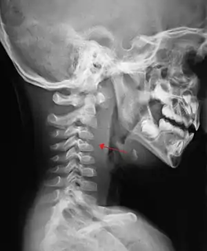
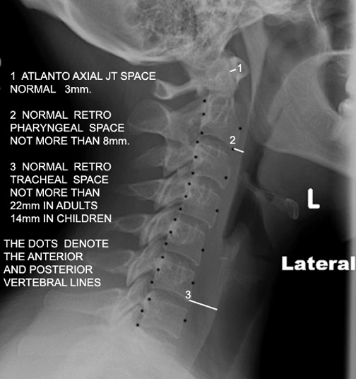
If more than half of the size of the C2 vertebra, it may indicate retropharyngeal abscess.[6]
Additional images
See also
References
![]() This article incorporates text in the public domain from page 390 of the 20th edition of Gray's Anatomy (1918)
This article incorporates text in the public domain from page 390 of the 20th edition of Gray's Anatomy (1918)
- Mnatsakanian A, Minutello K, Bordoni B (2022). "Anatomy, Head and Neck, Retropharyngeal Space". StatPearls. Treasure Island (FL): StatPearls Publishing. PMID 30725729. Retrieved 2022-07-24.
- Chong VF, Fan YF (October 2000). "Radiology of the Retropharyngeal Space". Clinical Radiology. 55 (10): 740–748. doi:10.1053/crad.2000.0510. PMID 11052873.
- "Spatium retropharyngeum". TheFreeDictionary.com. Retrieved 2022-07-24.
- Debnam JM, Guha-Thakurta N (December 2012). "Retropharyngeal and prevertebral spaces: anatomic imaging and diagnosis". Otolaryngologic Clinics of North America. 45 (6): 1293–1310. doi:10.1016/j.otc.2012.08.004. PMC 3994542. PMID 23153750.
- Jain H, Knorr TL, Sinha V (2022). Retropharyngeal Abscess. StatPearls. Treasure Island (FL): StatPearls Publishing. PMID 28722903. Retrieved 2022-07-24.
- Frank G, Shah SS, Catallozzi M, Zaoutis LB (1 June 2005). The Philadelphia guide: inpatient pediatrics. Lippincott Williams & Wilkins. pp. 181–. ISBN 978-1-4051-0428-9. Retrieved 26 May 2010.
External links
- Anatomy figure: 31:01-04 at Human Anatomy Online, SUNY Downstate Medical Center
- MedEd at Loyola Radio/curriculum/Bones/Image476a.jpg