Tuberculoma
A tuberculoma is a clinical manifestation of tuberculosis which conglomerates tubercles into a firm lump, and so can mimic cancer tumors of many types in medical imaging studies.[1][2] They often arise within individuals in whom a primary tuberculosis infection is not well controlled.[3] Since these are evolutions of primary complex, the tuberculomas may contain caseum or calcifications.
With the passage of time Mycobacterium tuberculosis can transform into crystals of calcium. These can affect any organ such as the brain,[4][5] intestine,[6][7][8] ovaries,[9][10] breast,[11][12][13] lungs,[14][15] esophagus,[16] pancreas,[17] bones,[18][19] and many others. Even with guideline-directed treatment they often persist for months to years.[3]
Epidemiology
Tuberculomas are most commonly seen in areas where tuberculosis is endemic. In these areas, tuberculomas can account for between 30%-50% of intracranial masses.[20][3] India and parts of Asia are two areas where tuberculomas have been noted to be particularly prevalent.[21] They occur most often as solitary lesions in young children.[3]
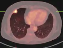
Pulmonary tuberculomas are among the most common benign nodules, with 5%-24% of all resected nodules being of tuberculous origin.[22] In areas of lower prevalence, such as the United States, they are most frequently seen in the setting of an acquired immunodeficiency.[23]
Signs and Symptoms
Symptoms are based on the location of the tuberculoma. Small, scattered lesions may be asymptomatic. Intracranial tuberculomas in children are often infratentorial, occurring near the cerebellum and base of the brain. In this population, symptoms such as headache, fever, focal neurologic findings and seizures have been seen[3] in addition to papilledema with or without meningitis.[21] When the size of a brainstem tuberculoma grows to the point of narrowing the fourth ventricle, obstructing hydrocephalus and its related symptoms can arise.[21] Rupture of tuberculomas adjacent to the arachnoid can lead to arachnoiditis.[23]
Imaging
The appearance of a tuberculoma on imaging can vary according to the composition and age of the mass. They may appear as either non-caseating or solidly caseating lesions.[21] Initially, tuberculomas appear hypodense on CT with significant surrounding edema.[23][3] The "target sign" is pathognomonic for tuberculoma on CT, with a nodular ring-enhancing mass and central calcification.[24][21] The characteristic ring-enhanced appearance is due to lack of blood supply in the central necrotic core that is visualized with injected contrast.[20] Sometimes a hypodense central area is seen instead of calcification.[25] When considering other potential intracranial masses in a differential diagnosis, such as cysticercosis, pyogenic abscess, and neoplastic lesions, tuberculoma can be identified by its larger size (>2 cm), edema, and irregular border.
MRI is another useful imaging modality for diagnosing and characterizing of tuberculomas, especially solid caseous necrosis in which 3 zones of varying intensity are seen.[20]
Treatment
Tuberculoma is commonly treated through the HRZE drug combination (Isoniazid, Rifampin, Pyrazinamide, Ethambutol) followed by maintenance therapy.[26] Per international guidelines, 9–12 months of medical management is standard.[21] While the majority of tuberculomas resolve in 12–24 months, in patients with multiple or larger lesions prolonged treatment extending beyond two years may be required. In some patients, the release of inflammatory mediators during treatment can cause a paradoxical worsening of symptoms that is treated with anti-inflammatory medications in addition to the standard anti-tuberculosis regimen.[20]
Exceptionally large tuberculomas, those exerting a mass effect on the brain, and those which fail to respond to medical management required surgical excision. In some cases, surgical excision is necessary for diagnosis as well as treatment.[3] When intracranial pressure rises in the setting of tuberculoma, removal is considered a surgical emergency.[20]
References
- Pitlik SD, Fainstein V, Bodey GP (May 1984). "Tuberculosis mimicking cancer--a reminder". The American Journal of Medicine. 76 (5): 822–5. doi:10.1016/0002-9343(84)90993-8. PMID 6720729.
- Vento S, Lanzafame M (June 2011). "Tuberculosis and cancer: a complex and dangerous liaison". The Lancet. Oncology. 12 (6): 520–2. doi:10.1016/S1470-2045(11)70105-X. PMID 21624773.
- Lloyd N. Friedman; Martin Dedicoat; P. D. O. Davies, eds. (2020). Clinical tuberculosis (Sixth ed.). Boca Raton, FL. ISBN 978-1-351-24998-0. OCLC 1145905400.
- Dennison P, Rajakaruna G (October 2006). "Cerebral tuberculoma". Thorax. 61 (10): 922. doi:10.1136/thx.2005.054932. PMC 2104774. PMID 17008487.
- Chatterjee S (October 2011). "Brain tuberculomas, tubercular meningitis, and post-tubercular hydrocephalus in children". Journal of Pediatric Neurosciences. 6 (Suppl 1): S96–S100. doi:10.4103/1817-1745.85725. PMC 3208909. PMID 22069437.
- Herrick FC (April 1925). "Tuberculoma of the Caecum: Hyperplastic Tuberculosis". Annals of Surgery. 81 (4): 801–20. doi:10.1097/00000658-192504000-00009. PMC 1399989. PMID 17865239.
- Chakravartty S, Chattopadhyay G, Ray D, Choudhury CR, Mandal S (2010). "Concomitant tuberculosis and carcinoma colon: coincidence or causal nexus?". Saudi Journal of Gastroenterology. 16 (4): 292–4. doi:10.4103/1319-3767.70619. PMC 2995101. PMID 20871197.
- Kushwaha JK, Sonkar AA, Saraf A, Singh D, Gupta R (September 2011). "Jejunal adenocarcinoma: an elusive diagnosis". Indian Journal of Surgical Oncology. 2 (3): 197–201. doi:10.1007/s13193-011-0101-7. PMC 3272177. PMID 22942611.
- Elmore RG, Li AJ (December 2007). "Peritoneal tuberculosis mimicking advanced-stage epithelial ovarian cancer". Obstetrics and Gynecology. 110 (6): 1417–9. doi:10.1097/01.AOG.0000295653.32975.4a. PMID 18055741.
- Rabesalama S, Mandeville K, Raherison R, Rakoto-Ratsimba H (2011). "Isolated ovarian tuberculosis mimicking ovarian carcinoma: case report and literature review". African Journal of Infectious Diseases. 5 (1): 7–10. doi:10.4314/ajid.v5i1.66508. PMC 3497843. PMID 23878702.
- Baharoon S (July 2008). "Tuberculosis of the breast". Annals of Thoracic Medicine. 3 (3): 110–4. doi:10.4103/1817-1737.41918. PMC 2700437. PMID 19561892.
- Sen M, Gorpelioglu C, Bozer M (2009). "Isolated primary breast tuberculosis: report of three cases and review of the literature". Clinics. 64 (6): 607–10. doi:10.1590/S1807-59322009000600019. PMC 2705158. PMID 19578668.
- Akçay MN, Sağlam L, Polat P, Erdoğan F, Albayrak Y, Povoski SP (June 2007). "Mammary tuberculosis -- importance of recognition and differentiation from that of a breast malignancy: report of three cases and review of the literature". World Journal of Surgical Oncology. 5: 67. doi:10.1186/1477-7819-5-67. PMC 1910599. PMID 17577397.
- Liang HY, Li XL, Yu XS, Guan P, Yin ZH, He QC, Zhou BS, et al. (December 2009). "Facts and fiction of the relationship between preexisting tuberculosis and lung cancer risk: a systematic review". International Journal of Cancer. 125 (12): 2936–44. doi:10.1002/ijc.24636. PMID 19521963. S2CID 21083607.
- Khan AN, Al-Jahdali HH, Allen CM, Irion KL, Al Ghanem S, Koteyar SS (April 2010). "The calcified lung nodule: What does it mean?". Annals of Thoracic Medicine. 5 (2): 67–79. doi:10.4103/1817-1737.62469. PMC 2883201. PMID 20582171.
- Patnayak R, Reddy MK, Parthasarathy S, Yootla M, Reddy V, Jena A (April 2008). "Unusual presentation of esophageal tuberculosis mimicking malignancy". Saudi Journal of Gastroenterology. 14 (2): 103–4. doi:10.4103/1319-3767.39632. PMC 2702907. PMID 19568514.
- Saluja SS, Ray S, Pal S, Kukeraja M, Srivastava DN, Sahni P, Chattopadhyay TK (June 2007). "Hepatobiliary and pancreatic tuberculosis: a two decade experience". BMC Surgery. 7 (1): 10. doi:10.1186/1471-2482-7-10. PMC 1925057. PMID 17588265.
- Herzog A (September 2009). "Dangerous errors in the diagnosis and treatment of bony tuberculosis". Deutsches Ärzteblatt International. 106 (36): 573–7. doi:10.3238/arztebl.2009.0573. PMC 2770211. PMID 19890413.
- Dhillon MS, Aggarwal S, Prabhakar S, Bachhal V (March 2012). "Tuberculosis of the foot: An osteolytic variety". Indian Journal of Orthopaedics. 46 (2): 206–11. doi:10.4103/0019-5413.93683. PMC 3308663. PMID 22448060.
- Perez-Malagon, Carlos David; Barrera-Rodriguez, Raul; Lopez-Gonzalez, Miguel A.; Alva-Lopez, Luis F. (December 2021). "Diagnostic and Neurological Overview of Brain Tuberculomas: A Review of Literature". Cureus. 13 (12): e20133. doi:10.7759/cureus.20133. ISSN 2168-8184. PMC 8648135. PMID 34900500.
- Gupta, Monica; Munakomi, Sunil (2022), "CNS Tuberculosis", StatPearls, Treasure Island (FL): StatPearls Publishing, PMID 36256788, retrieved 2022-10-31
- Lee, H. S.; Oh, J. Y.; Lee, J. H.; Yoo, C. G.; Lee, C. T.; Kim, Y. W.; Han, S. K.; Shim, Y. S.; Yim, J. J. (March 2004). "Response of pulmonary tuberculomas to anti-tuberculous treatment". The European Respiratory Journal. 23 (3): 452–455. doi:10.1183/09031936.04.00087304. ISSN 0903-1936. PMID 15065838.
- Martin A. Samuels; Steven K. Feske, eds. (2003). Office practice of neurology (2nd ed.). Philadelphia: Churchill Livingstone. ISBN 978-0-7020-3588-3. OCLC 324998368.
- Kateryna Kon; Mahendra Rai, eds. (2018). The microbiology of central nervous system infections. London. ISBN 978-0-12-813807-6. OCLC 1023628139.
- Michael J. Aminoff; Scott Andrew Josephson, eds. (2021). Aminoff's neurology and general medicine (Sixth ed.). London. ISBN 978-0-12-819307-5. OCLC 1235762322.
- Monteiro R, Carneiro JC, Costa C, Duarte R (2013). "Cerebral tuberculomas - A clinical challenge". Respiratory Medicine Case Reports. 9: 34–7. doi:10.1016/j.rmcr.2013.04.003. PMC 3949551. PMID 26029627.