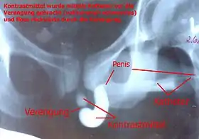
Retrograde urethrogram
A retrograde urethrography[1] is a routine radiologic procedure (most typically in males) used to image the integrity of the urethra. Hence a retrograde urethrogram is essential for diagnosis of urethral injury, or urethral stricture.[2][3]
| Retrograde urethrography | |
|---|---|
 Urethrogram showing an urethra stricture in a man. | |
| ICD-9 | 87.76 |
| OPS-301 code | 3-13g |
Uses
Some indications for retrograde urethrogram are: urethral stricture, urethral trauma, urethral fistula and congenital urethral abnormalities.[4] There is no absolute contraindication for retrograde urethrogram. There are several relative contraindications such as: allergy to contrast agents, acute urinary tract infection, and recent instrumentation of urethra.[5]
Procedure
A low osmolar contrast agent with concentration of 200 to 300 mg per ml with volume of 20 ml can be used in this study. Warming the contrast medium before infusion into the urethra can help to reduce the chance of getting spasm of external urethral sphincter.[4]
The subject lie down on supine position. An 8 Fr Foley catheter is connected to a 50 ml syringe. The syringe is flushed to remove any air bubbles within the Foley catheter and the syringe. The tip of the catheter is then inserted into the urethra using aseptic technique until it park inside the navicular fossa. Fossa navicularis is located just a short distance proximal to urethral meatus within the glans penis. The balloon of the Foley catheter is then inflated with 2 to 3 ml of water to anchor the catheter and occlude the meatus, thus prevent contrast from leaking out from the penis. Contrast is then injected from the syringe with fluoroscopy to visualise the flow of contrast within the penis. The catheter is gentlely pulled to straighten the penis over the leg of the same side to prevent the overlapping of any pathology in the posterior urethrae. Spot images are taken at 30 to 45 degrees to visualise the entire spongy urethra (penile urethra).[4]
If there is no contraindication to full urinary catheterisation such as false passage or stricture, the urinary catheter should be inserted until urinary bladder to perform voiding cystourethrography to visualise the prostatic urethra and membranous urethra. Filling up the bladder with contrast without full catheterisation (the end of catheter inside the urethra) is also possible if the subject is able to relax the bladder neck to allow contrast to follow into the bladder.[4]
If a urethral injury is suspected, a retrograde urethrography should be performed before attempting to place a Foley catheter into the bladder. If there is a urethral disruption, a suprapubic catheter should be placed.
Complications
Among the possible complications are: urinary tract infection, urethral trauma, and intravasation of contrast medium (contrast going into blood vessels) if excessive pressure is used to overcome a stricture.[4]
See also
- Retrograde ureteral, an intervention used to remove kidney stones
References
- Shetty, Aditya. "Urethrography | Radiology Reference Article | Radiopaedia.org". Radiopaedia. Retrieved 2021-11-10.
- El-Ghar MA, Osman Y, Elbaz E, Refiae H, El-Diasty T (July 2009). "MR urethrogram versus combined retrograde urethrogram and sonourethrography in diagnosis of urethral stricture". Eur J Radiol. 74 (3): e193–e198. doi:10.1016/j.ejrad.2009.06.008. PMID 19608363.
- Maciejewski, Conrad; Rourke, Keith (2015-12-02). "Imaging of urethral stricture disease". Translational Andrology and Urology. 4 (1): 2–9. doi:10.3978/j.issn.2223-4683.2015.02.03. ISSN 2223-4691. PMC 4708283. PMID 26816803.
- Watson N, Jones H (2018). Chapman and Nakielny's Guide to Radiological Procedures. Elsevier. pp. 140–141. ISBN 9780702071669.
- Hota, Parta; Patel, Tejas; Patel, Harshad; et al. (February 2020). "Post-traumatic Retrograde Urethrography: A Review of Acute Findings and Chronic Complications". Applied Radiology. 49 (1): 24–31.
External links
- New England Journal of Medicine procedure videos: Male Urethral catheterization
- Ohio State University Patient Education Materials: Retrograde Urethrogram