Viral envelope
A viral envelope is the outermost layer of many types of viruses.[1] It protects the genetic material in their life cycle when traveling between host cells. Not all viruses have envelopes.

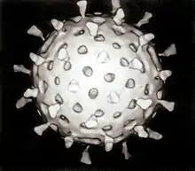
The envelopes are typically derived from portions of the host cell membranes (phospholipids and proteins), but include some viral glycoproteins. Being made up mostly of host membrane, the viral envelope can also have the proteins associated with the host cell within their membrane after budding.[2] They may help viruses avoid the host immune system. Glycoproteins on the surface of the envelope serve to identify and bind to receptor sites on the host's membrane. The particular set of viral proteins are engaged in a series of structural changes. When these changes are set/finished, there is then and only then, fusion with the host membrane.[3] These glycoproteins mediate the interaction between virion and host cell, typically initiating the fusion between the viral envelope and the host's cellular membrane.[4] In some cases, the virus with an envelope will form an endosome within the host cell.[5] There are three main types of viral glycoproteins: Envelope proteins, membrane proteins, and spike proteins (E, M, and S).[6] The viral envelope then fuses with the host's membrane, allowing the capsid and viral genome to enter and infect the host.
All enveloped viruses also have a capsid, another protein layer, between the envelope and the genome.[1] The capsid, having a focused role of protecting the genome in addition to immune recognition evasion.[7] The viral capsid is known for its protection of RNA before it is inserted into the host cell, unlike the viral envelope which protects the protein capsid.[8]
The cell from which a virus buds often dies or is weakened, and sheds more viral particles for an extended period. The lipid bilayer envelope of these viruses is relatively sensitive to desiccation, heat, and amphiphiles such as soap and detergents, therefore these viruses are easier to sterilize than non-enveloped viruses, have limited survival outside host environments, and typically must transfer directly from host to host. Viral envelope persistence, wither it be enveloped or naked, are a factor in determining longevity of a virus on inanimate surfaces.[9] Enveloped viruses possess great adaptability and can change in a short time in order to evade the immune system. Enveloped viruses can cause persistent infections.
Vaccination against enveloped viruses can function by neutralizing the glycoprotein activity with antibodies.[10]
Examples of enveloped viruses
The following are some examples of enveloped viruses:
- DNA viruses
- Herpesviruses
- Poxviruses
- Hepadnaviruses
- Asfarviridae
- RNA viruses
- Flaviviruses
- Alphaviruses
- Togaviruses
- Coronaviruses
- Hepatitis D
- Orthomyxoviruses
- Paramyxoviruses
- Rhabdovirus[11]
- Bunyaviruses
- Filoviruses
- Retroviruses
Examples of non-enveloped viruses
The following are some examples of viruses without envelopes:
- DNA viruses
- Adenoviruses
- Parvoviruses
- Polyomaviruses
- Anelloviruses
- RNA viruses
- Caliciviruses
- Picornaviruses
- Reoviruses
- Astroviruses
- Hepeviridae
See also
- Bacterial capsule
- Cell envelope
References
- HURLBERT, RONALD E. Fundamentals of Microbiology 102. Chapter #11: Viruses. Archived from the original on 2008-11-10. Retrieved 2008-11-07.
- Gelderblom HR. Structure and Classification of Viruses. In: Baron S, editor. Medical Microbiology. 4th edition. Galveston (TX): University of Texas Medical Branch at Galveston; 1996. Chapter 41. Available from: https://www.ncbi.nlm.nih.gov/books/NBK8174/
- Benhaim, Mark A.; Lee, Kelly K. (2020-04-08). "New Biophysical Approaches Reveal the Dynamics and Mechanics of Type I Viral Fusion Machinery and Their Interplay with Membranes". Viruses. 12 (4): E413. doi:10.3390/v12040413. ISSN 1999-4915. PMC 7232462. PMID 32276357.
- Navaratnarajah, C.K. et al. “Assembly of Viruses: Enveloped Particles.” Encyclopedia of Virology (2008): 193–200. doi:10.1016/B978-012374410-4.00667-1
- White, Judith M, and Gary R Whittaker. “Fusion of Enveloped Viruses in Endosomes.” Traffic (Copenhagen, Denmark) vol. 17,6 (2016): 593-614. doi:10.1111/tra.12389
- Banerjee, Nilotpal, and Sumi Mukhopadhyay. “Viral glycoproteins: biological role and application in diagnosis.” Virusdisease vol. 27,1 (2016): 1-11. doi:10.1007/s13337-015-0293-5
- Stuart, David I.; Ren, Jingshan; Wang, Xiangxi; Rao, Zihe; Fry, Elizabeth E. (May 2019). "Hepatitis A Virus Capsid Structure". Cold Spring Harbor Perspectives in Medicine. 9 (5): a031807. doi:10.1101/cshperspect.a031807. ISSN 2157-1422. PMC 6496327. PMID 30037986.
- Cliver, Dean O. (2009). "Capsid and Infectivity in Virus Detection". Food and Environmental Virology. 1 (3): 123–128. doi:10.1007/s12560-009-9020-y. ISSN 1867-0334. PMC 2837222. PMID 20234879.
- Firquet, Swan; Beaujard, Sophie; Lobert, Pierre-Emmanuel; Sané, Famara; Caloone, Delphine; Izard, Daniel; Hober, Didier (June 2015). "Survival of Enveloped and Non-Enveloped Viruses on Inanimate Surfaces". Microbes and Environments. 30 (2): 140–144. doi:10.1264/jsme2.ME14145. ISSN 1342-6311. PMC 4462923. PMID 25843687.
- Rey, Felix A.; Lok, Shee-Mei (8 March 2018). "Common Features of Enveloped Viruses and Implications for Immunogen Design for Next-Generation Vaccines". Cell. 172 (6): 1319–1334. doi:10.1016/j.cell.2018.02.054. PMC 7112304. PMID 29522750.
- "The Rabies Virus". CDC. Archived from the original on 2017-01-18. Retrieved 2008-11-07.
External links
- "Virus Structure". Molecular Expressions: Images from the Microscope. Retrieved 2007-06-27.
| ||
| Components |
|  |
| Viral life cycle |
| |
| Genetics |
| |
| By host |
| |
| Other |
| |
| ||