Animal Reproduction and Development
Most animals are diploid organisms (their body, or somatic, cells are diploid) with haploid reproductive (gamete) cells produced through meiosis. The majority of animals undergo sexual reproduction. This fact distinguishes animals from fungi, protists, and bacteria where asexual reproduction is common or exclusive. However, a few groups, such as cnidarians, flatworms, and roundworms, undergo asexual reproduction, although nearly all of those animals also have a sexual phase to their life cycle.
Processes of Animal Reproduction and Embryonic Development
During sexual reproduction, the haploid gametes of the male and female individuals of a species combine in a process called fertilization. Typically, the small, motile male sperm fertilizes the much larger, sessile female egg. This process produces a diploid fertilized egg called a zygote.
Some animal species (including sea stars and sea anemones, as well as some insects, reptiles, and fish) are capable of asexual reproduction. The most common forms of asexual reproduction for stationary aquatic animals include budding and fragmentation where part of a parent individual can separate and grow into a new individual. In contrast, a form of asexual reproduction found in certain insects and vertebrates is called parthenogenesis where unfertilized eggs can develop into new offspring. This type of parthenogenesis in insects is called haplodiploidy and results in male offspring. These types of asexual reproduction produce genetically identical offspring, which is disadvantageous from the perspective of evolutionary adaptability because of the potential buildup of deleterious mutations. However, for animals that are limited in their capacity to attract mates, asexual reproduction can ensure genetic propagation.
After fertilization, a series of developmental stages occur during which primary germ layers are established and reorganize to form an embryo. During this process, animal tissues begin to specialize and organize into organs and organ systems, determining their future morphology and physiology. Some animals, such as grasshoppers, undergo incomplete metamorphosis, in which the young resemble the adult. Other animals, such as some insects, undergo complete metamorphosis where individuals enter one or more larval stages that may differ in structure and function from the adult . In complete metamorphosis, the young and the adult may have different diets, limiting competition for food between them. Regardless of whether a species undergoes complete or incomplete metamorphosis, the series of developmental stages of the embryo remains largely the same for most members of the animal kingdom.

Incomplete and complete metamorphosis
(a) The grasshopper undergoes incomplete metamorphosis. (b) The butterfly undergoes complete metamorphosis.
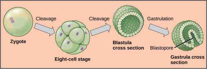
The process of animal development begins with the cleavage, or series of mitotic cell divisions, of the zygote . Three cell divisions transform the single-celled zygote into an eight-celled structure. After further cell division and rearrangement of existing cells, a 6–32-celled hollow structure called a blastula is formed. Next, the blastula undergoes further cell division and cellular rearrangement during a process called gastrulation. This leads to the formation of the next developmental stage, the gastrula, in which the future digestive cavity is formed. Different cell layers (called germ layers) are formed during gastrulation. These germ layers are programed to develop into certain tissue types, organs, and organ systems during a process called organogenesis.

Embryonic development
During embryonic development, the zygote undergoes a series of mitotic cell divisions, or cleavages, to form an eight-cell stage, then a hollow blastula. During a process called gastrulation, the blastula folds inward to form a cavity in the gastrula.
The Role of Homeobox (Hox) Genes in Animal Development
Since the early 19th century, scientists have observed that many animals, from the very simple to the complex, shared similar embryonic morphology and development. Surprisingly, a human embryo and a frog embryo, at a certain stage of embryonic development, appear remarkably similar. For a long time, scientists did not understand why so many animal species looked similar during embryonic development, but were very different as adults. Near the end of the 20th century, a particular class of genes that dictate developmental direction was discovered. These genes that determine animal structure are called "homeotic genes." They contain DNA sequences called homeoboxes, with specific sequences referred to as Hox genes. This family of genes is responsible for determining the general body plan: the number of body segments of an animal, the number and placement of appendages, and animal head-tail directionality. The first Hox genes to be sequenced were those from the fruit fly (Drosophila melanogaster). A single Hox mutation in the fruit fly can result in an extra pair of wings or even appendages growing from the "wrong" body part.
There are many genes that play roles in the morphological development of an animal, but Hox genes are so powerful because they can turn on or off large numbers of other genes. Hox genes do this by coding transcription factors that control the expression of numerous other genes. Hox genes are homologous in the animal kingdom: the genetic sequences and their positions on chromosomes are remarkably similar across most animals (e.g., worms, flies, mice, humans) because of their presence in a common ancestor . Hox genes have undergone at least two duplication events during animal evolution: the additional genes allowed more complex body types to evolve.

Hox genes
Hox genes are highly-conserved genes encoding transcription factors that determine the course of embryonic development in animals. In vertebrates, the genes have been duplicated into four clusters: Hox-A, Hox-B, Hox-C, and Hox-D. Genes within these clusters are expressed in certain body segments at certain stages of development. Shown here is the homology between Hox genes in mice and humans. Note how Hox gene expression, as indicated with orange, pink, blue, and green shading, occurs in the same body segments in both the mouse and the human.