Concept
Version 12
Created by Boundless
Trachea

Histology of the Trachea
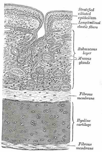
A cross section of the trachea, showing the hyaline cartilage, mucus glands, and ciliated epithelium.
A cross section of the trachea, showing the hyaline cartilage, mucus glands, and ciliated epithelium. The hyaline cartilage is wedged between two fibrous membranes. The submucous layer contains the mucous glands. The stratified ciliated ephithelium sits above it all, cushioned by longitudinal elastic fibers.
Source
Boundless vets and curates high-quality, openly licensed content from around the Internet. This particular resource used the following sources:
"Gray964.png."
https://upload.wikimedia.org/wikipedia/commons/f/f7/Gray964.png
Wikipedia Commons
CC BY-SA 3.0.