Voluntary respiration is any type of respiration that is under conscious control. Voluntary respiration is important for the higher functions that involve air supply, such as voice control or blowing out candles. Similarly to how involuntary respiration's lower functions are controlled by the lower brain, voluntary respiration's higher functions are controlled by the upper brain, namely parts of the cerebral cortex.
The Motor Cortex
The primary motor cortex is the neural center for voluntary respiratory control. More broadly, the motor cortex is responsible for initiating any voluntary muscular movement.
The processes that drive its functions aren't fully understood, but it works by sending signals to the spinal cord, which sends signals to the muscles it controls, such as the diaphragm and the accessory muscles for respiration. This neural pathway is called the ascending respiratory pathway.
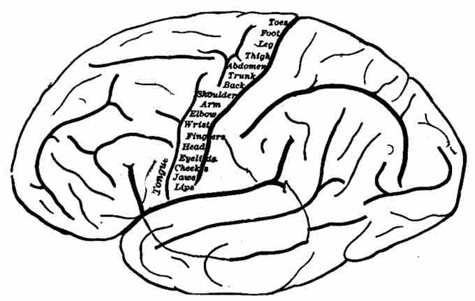
Different parts of the cerebral cortex control different forms of voluntary respiration. Initiation of the voluntary contraction and relaxation of the internal and external intercostal muscles takes place in the superior portion of the primary motor cortex.
The center for diaphragm control is posterior to the location of thoracic control (within the superior portion of the primary motor cortex). The inferior portion of the primary motor cortex may be involved in controlled exhalation.
Activity has also been seen within the supplementary motor area and the premotor cortex during voluntary respiration. This is most likely due to the focus and mental preparation of the voluntary muscular movement that occurs when one decides to initiate that muscle movement.
Note that voluntary respiratory nerve signals in the ascending respiratory pathway can be overridden by chemoreceptor signals from involuntary respiration. Additionally, other structures may override voluntary respiratory signals, such as the activity of limbic center structures like the hypothalamus.
During periods of perceived danger or emotional stress, signals from the hypothalamus take over the respiratory signals and increase the respiratory rate to facilitate the fight or flight response.

Topography of the primary motor cortex
Topography of the primary motor cortex, on an outline drawing of the human brain. Each part of the primary motor cortex controls a different part of the body.
Nerves Used in Respiration
There are several nerves responsible for the muscular functions involved in respiration. There are three types of important respiratory nerves:
- The phrenic nerves: The nerves that stimulate the activity of the diaphragm. They are composed of two nerves, the right and left phrenic nerve, which pass through the right and left side of the heart respectively. They are autonomic nerves.
- The vagus nerve: Innervates the diaphragm as well as movements in the larynx and pharynx. It also provides parasympathetic stimulation for the heart and the digestive system. It is a major autonomic nerve.
- The posterior thoracic nerves: These nerves stimulate the intercostal muscles located around the pleura. They are considered to be part of a larger group of intercostal nerves that stimulate regions across the thorax and abdomen. They are somatic nerves.
These three types of nerves continue the signal of the ascending respiratory pathway from the spinal cord to stimulate the muscles that perform the movements needed for respiration.
Damage to any of these three respiratory nerves can cause severe problems, such as diaphragm paralysis if the phrenic nerves are damaged. Less severe damage can cause irritation to the phrenic or vagus nerves, which can result in hiccups.