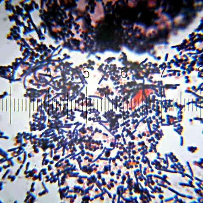
Gram-positive bacteria are stained dark blue or violet by Gram staining . While Gram staining is a valuable diagnostic tool in both clinical and research settings, not all bacteria can be definitively classified by this technique, thus forming Gram-variable and Gram-indeterminate groups as well.

Gram-positive bacteria
These bacteria stain violet by Gram staining.
It is based on the chemical and physical properties of their cell walls. Primarily, it detects peptidoglycan, which is present in a thick layer in Gram-positive bacteria. A Gram-positive results in a purple/blue color while a Gram-negative results in a pink/red color. The Gram stain is almost always the first step in the identification of a bacterial organism, and is the default stain performed by laboratories over a sample when no specific culture is referred.
In Gram-positive bacteria, the cell wall is thick (15-80 nanometers), and consists of several layers of peptidoglycan. They lack the outer membrane envelope found in Gram-negative bacteria. Running perpendicular to the peptidoglycan sheets is a group of molecules called teichoic acids, which are unique to the Gram-positive cell wall. Teichoic acids are linear polymers of polyglycerol or polyribitol substituted with phosphates and a few amino acids and sugars.
The teichoic acid polymers are occasionally anchored to the plasma membrane (called lipoteichoic acid, LTA), and apparently directed outward at right angles to the layers of peptidoglycan. Teichoic acids give the Gram-positive cell wall an overall negative charge due to the presence of phosphodiester bonds between teichoic acid monomers. The functions of teichoic acid are not fully known but it is believed to serve as a chelating agent and means of adherence for the bacteria. These are essential to the viability of Gram-positive bacteria in the environment and provide chemical and physical protection.
One idea is that they provide a channel of regularly-oriented, negative charges for threading positively-charged substances through the complicated peptidoglycan network. Another theory is that teichoic acids are in some way involved in the regulation and assembly of muramic acid sub-units on the outside of the plasma membrane.
There are instances, particularly in the streptococci, wherein teichoic acids have been implicated in the adherence of the bacteria to tissue surfaces and are thought to contribute to the pathogenicity of Gram-positive bacteria.