Lymphogranuloma venereum (LGV) is a sexually transmitted disease which was considered rare in the developed world until about a decade ago. LGV is an infection of the lymph nodes. The infectious agent enters the body through breaks in the skin or through the epithelial layer of mucous membranes.
Infectious Agents
The infectious agents are a few serovars of Chlamydia trachomatis : L1, L2, L2a, L2b and L3.

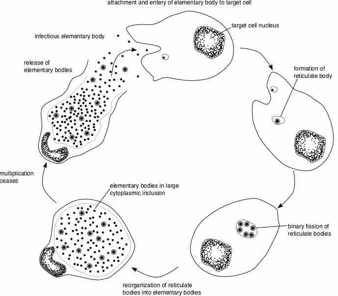
Chlamydiae Life Cycle
Basic diagram of the life cycle of the Chlamydiae. The infectious agents of Lymphogranuloma venereum are a few serovars of Chlamydia trachomatis: L1, L2, L2a, L2b and L3.
Symptoms and Diagnosis
The general symptoms may include fever, malaise and decreased appetite. The disease progresses in stages. In the primary stage, symptoms appear within days after infection. The first symptom is usually painless ulcers at the contact area.The secondary stage can manifest from days to months later. The infectious agent spreads to the lymph nodes through the lymphatic drainage pathways, causing inflammation of the lymph nodes and lymphatic channels. In males with genital infection, these symptoms will usually be in the inguinal or/and femoral areas. In women, an inflammation of the cervix, the fallopian tubes or/and peritonitis may appear as well as inflammation and infection of the lymphatic system. If the infection started in the anal area, it may cause inflammation of the rectum or the colonic mucosa, presenting with symptoms such as anorectal pain, discharge, abdominal cramps and diarrhea.
The enlarged lymph nodes are called buboes and are painful, inflamed and can fixate to the skin. These changes can further progress to necrosis, abscesses and fistulas. As healing starts, fibrosis may occur in the inflamed areas and cause obstruction of the lymphatic system and edema. The fibrosis and edema are considered the third stage of LGV and are mainly permanent. Diagnosis is made after serological analysis and exclusion of other reasons for genital ulcers and lymphatic issues. Culturing is also used for identification of serotypes. Other tests include direct fluorescent antibody analysis (DFA) and PCR tests.
Treatment
Treatment is performed with antibiotics, usually tetracycline, doxycycline or erythromycin. Sometimes drainage of the buboes or abscesses is performed as well. Prognosis is best if treatment starts early in the infection process. Severe complications include bowel obstruction or perforation, which can lead to death.