Microorganisms are very diverse. They include bacteria, fungi, algae, and protozoa; microscopic plants (green algae); and animals such as rotifers and planarians. Most microorganisms are unicellular (single-celled), but this is not universal.
Single-celled microorganisms were the first forms of life to develop on earth, approximately 3 billion–4 billion years ago. Further evolution was slow, and for about 3 billion years in the Precambrian eon, all organisms were microscopic. So, for most of the history of life on earth the only forms of life were microorganisms. Bacteria, algae, and fungi have been identified in amber that is 220 million years old, which shows that the morphology of microorganisms has changed little since the Triassic period. When at the end of the 19th century information began to accumulate about the diversity within the bacterial world, scientists started to include the bacteria in phylogenetic schemes to explain how life on earth may have developed. Some of the early phylogenetic trees of the prokaryote world were morphology-based. Others were based on the then-current ideas on the presumed conditions on our planet at the time that life first developed.
Microorganisms tend to have a relatively rapid evolution. Most microorganisms can reproduce rapidly, and microbes such as bacteria can also freely exchange genes through conjugation, transformation, and transduction, even between widely-divergent species. This horizontal gene transfer, coupled with a high mutation rate and many other means of genetic variation, allows microorganisms to swiftly evolve (via natural selection) to survive in new environments and respond to environmental stresses.
The relationship between the three domains (Bacteria, Archaea, and Eukaryota) is of central importance for understanding the origin of life. Most of the metabolic pathways, which comprise the majority of an organism's genes, are common between Archaea and Bacteria, while most genes involved in genome expression are common between Archaea and Eukarya. Within prokaryotes, archaeal cell structure is most similar to that of Gram-positive bacteria.
Phenotypic Methods of Classifying and Identifying Microorganisms
Classification seeks to describe the diversity of bacterial species by naming and grouping organisms based on similarities. Microorganisms can be classified on the basis of cell structure , cellular metabolism, or on differences in cell components such as DNA, fatty acids, pigments, antigens, and quinones.

Bacterial Morphology
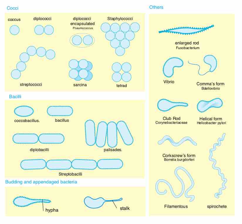
Basic morphological differences between bacteria. The most often found forms and their associations.
There are some basic differences between Bacteria, Archaea, and Eukaryotes in cell morphology and structure which aid in phenotypic classification and identification:

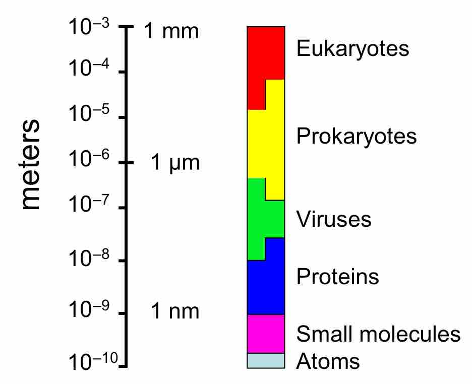
The relative sizes of prokaryotic cells
Relative scales of eukaryotes, prokaryotes, viruses, proteins and atoms (logarithmic scale).
- Bacteria: lack membrane-bound organelles and can function and reproduce as individual cells, but often aggregate in multicellular colonies. Their genome is usually a single loop of DNA, although they can also harbor small pieces of DNA called plasmids. These plasmids can be transferred between cells through bacterial conjugation. Bacteria are surrounded by a cell wall, which provides strength and rigidity to their cells.
- Archaea: In the past, the differences between bacteria and archaea were not recognized and archaea were classified with bacteria as part of the kingdom Monera. Archaea are also single-celled organisms that lack nuclei. Archaea in fact differ from bacteria in both their genetics and biochemistry. While bacterial cell membranes are made from phosphoglycerides with ester bonds, archaean membranes are made of ether lipids.
- Eukaryotes: Unlike bacteria and archaea, eukaryotes contain organelles such as the cell nucleus, the Golgi apparatus, and mitochondria in their cells. Like bacteria, plant cells have cell walls and contain organelles such as chloroplasts in addition to the organelles in other eukaryotes.
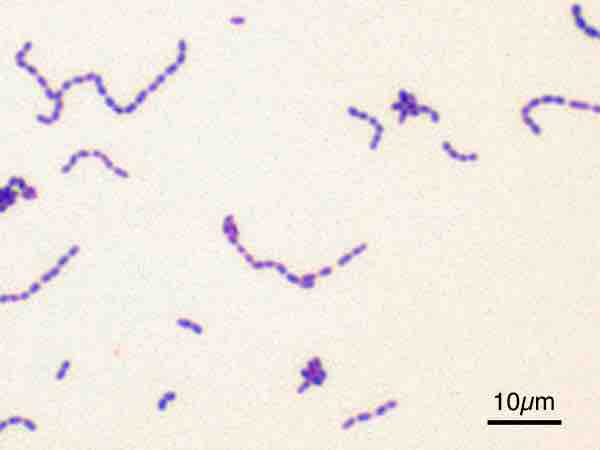
The Gram stain, developed in 1884 by Hans Christian Gram, characterizes bacteria based on the structural characteristics of their cell walls. The thick layers of peptidoglycan in the "Gram-positive" cell wall stain purple , while the thin "Gram-negative" cell wall appears pink. By combining morphology and Gram-staining, most bacteria can be classified as belonging to one of four groups (Gram-positive cocci, Gram-positive bacilli, Gram-negative cocci, and Gram-negative bacilli). Some organisms are best identified by stains other than the Gram stain, particularly mycobacteria or Nocardia, which show acid-fastness on Ziehl–Neelsen or similar stains. Other organisms may need to be identified by their growth in special media, or by other techniques, such as serology.

Gram-positive bacteria
Streptococcus mutans visualized with a Gram stain.
While these schemes allowed the identification and classification of bacterial strains, it was unclear whether these differences represented variation between distinct species or between strains of the same species. This uncertainty was due to the lack of distinct structures in most bacteria, as well as lateral gene transfer between unrelated species. Due to lateral gene transfer, some closely related bacteria can have very different morphologies and metabolisms. To overcome this uncertainty, modern bacterial classification emphasizes molecular systematics, using genetic techniques such as guanine cytosine ratio determination, genome-genome hybridization, as well as sequencing genes that have not undergone extensive lateral gene transfer, such as the rRNA gene.