The human visual system gives our bodies the ability to see our physical environment. The system requires communication between its major sensory organ (the eye) and the core of the central nervous system (the brain) to interpret external stimuli (light waves) as images. Humans are highly visual creatures compared to many other animals which rely more on smell or hearing, and over our evolutionary history we have developed an incredibly complex sight system.
Sensory Organs
Vision depends mainly on one sensory organ—the eye. Eye constructions vary in complexity depending on the needs of the organism. The human eye is one of the most complicated structures on earth, and it requires many components to allow our advanced visual capabilities. The eye has three major layers:
- the sclera, which maintains, protects, and supports the shape of the eye and includes the cornea;
- the choroid, which provides oxygen and nourishment to the eye and includes the pupil, iris, and lens; and
- the retina, which allows us to piece images together and includes cones and rods.
The Process of Sight
All vision is based on the perception of electromagnetic rays. These rays pass through the cornea in the form of light; the cornea focuses the rays as they enter the eye through the pupil, the black aperture at the front of the eye. The pupil acts as a gatekeeper, allowing as much or as little light to enter as is necessary to see an image properly. The pigmented area around the pupil is the iris. Along with supplying a person's eye color, the iris is responsible for acting as the pupil's stop, or sphincter. Two layers of iris muscles contract or dilate the pupil to change the amount of light that enters the eye. Behind the pupil is the lens, which is similar in shape and function to a camera lens. Together with the cornea, the lens adjusts the focal length of the image being seen onto the back of the eye, the retina. Visual reception occurs at the retina where photoreceptor cells called cones and rods give an image color and shadow. The image is transduced into neural impulses and then transferred through the optic nerve to the rest of the brain for processing. The visual cortex in the brain interprets the image to extract form, meaning, memory, and context.
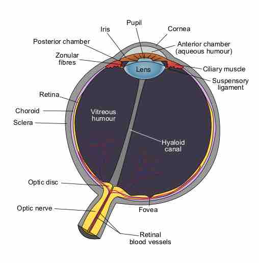
Anatomy of the human eye
A cross-section of the human eye with its component pieces labeled. Clockwise from left: Optic nerve, optic disc, sclera, choroid, retina, zonular fibers, posterior chamber, iris, pupil, cornea, aqueous humor, ciliary muscle, suspensory ligament, fovea, retinal blood vessels. In center: Vitreous humour, hyaloid canal, lens.
The left hemisphere of the brain controls the motor functions of the right half of the body, and vice versa; the same is true of vision. The left hemisphere of the brain processes visual images from the right-hand side of space, or the right visual field, and the right hemisphere processes visual images from the left-hand side of space, or the left visual field. The optic chiasm is a complicated crossover of optic nerve fibers behind the eyes at the bottom of the brain, allowing the right eye to "wire" to the left neural hemisphere and the left eye to "wire" to the right hemisphere. This allows the visual cortex to receive the same visual field from both eyes.
Color Vision
Human beings are capable of highly complex vision that allows us to perceive colors and depth in intricate detail. Visual stimulus transduction happens in the retina. Photoreceptor cells found in this region have the specialized capability of phototransduction, or the ability to convert light into electrical signals. There are two types of these photoreceptor cells: rods, which are responsible for scotopic vision (night vision), and cones, which are responsible for photopic vision (daytime vision).
Generally speaking, cones are for color vision and rods are for shadows and light differences. The front of your eye has many more cones than rods, while the sides have more rods than cones; for this reason, your peripheral vision is sharper than your direct vision in the darkness, but your peripheral vision is also in black and white.
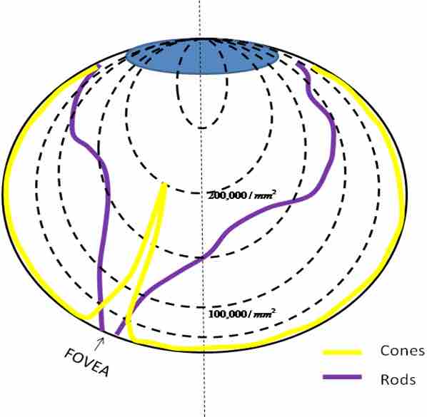
Cones and Rods
This density map shows the retina, which is made up of cones and rods. Cones perceive color and rods perceive shadow in images. In the fovea, which is responsible for sharp central vision, there is huge density of cones but no rods.
Color vision is a critical component of human vision and plays an important role in both perception and communication. Color sensors are found within cones, which respond to relatively broad color bands in the three basic regions of red, green, and blue (RGB). Any colors in between these three are perceived as different linear combinations of RGB. The eye is much more sensitive to overall light and color intensity than changes in the color itself. Colors have three attributes: brightness, based on luminance and reflectivity; saturation, based on the amount of white present; and hue, based on color combinations. Sophisticated combinations of these receptors signals are transduced into chemical and electrical signals, which are sent to the brain for the dynamic process of color perception.
Depth Perception
Depth perception refers to our ability to see the world in three dimensions. With this ability, we can interact with the physical world by accurately gauging the distance to a given object. While depth perception is often attributed to binocular vision (vision from two eyes), it also relies heavily on monocular cues (cues from only one eye) to function properly. These cues range from the convergence of our eyes and accommodation of the lens to optical flow and motion.