


Microsporidiosis
 ShareCompartir
[Anncaliia spp.] [Encephalitozoon cuniculi] [Encephalitozoon hellem] [Encephalitozoon intestinalis (syn. Septata intestinalis)] [Tubulinosema acridophagus] [Enterocytozoon bieneusi] [Nosema spp.] [Pleistophora sp.] [Trachipleistophora spp.] [Vittaforma corneae (syn. Nosema corneum)]
Causal Agents
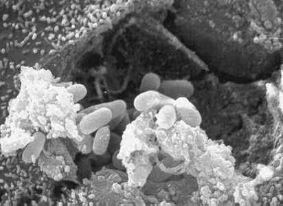
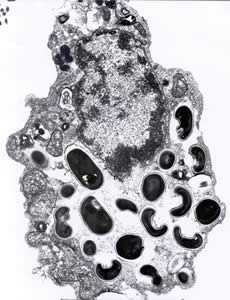
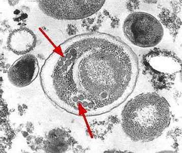
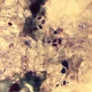
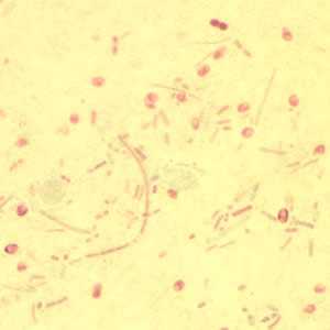
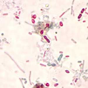
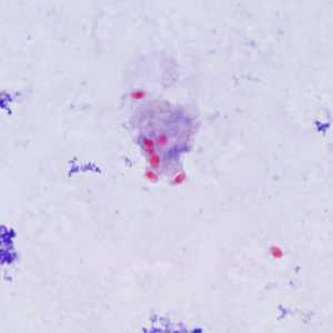
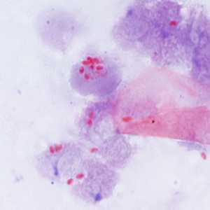
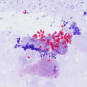
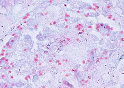
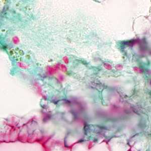
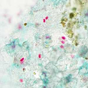
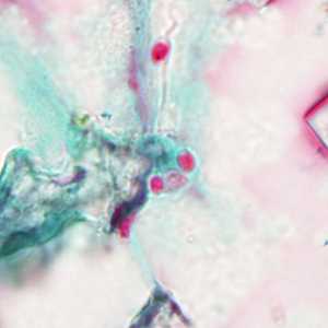
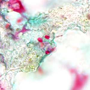
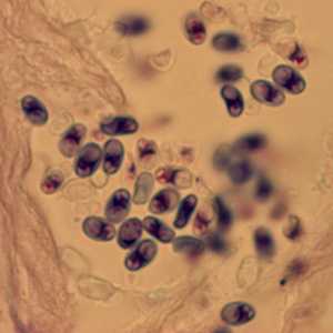
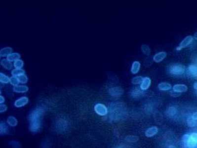
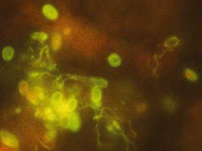
The microsporidia are a group of obligate intracellular parasitic fungi. Historically, they have been treated among the protozoa, and as such are often still managed by diagnostic parasitology laboratories. To date, more than 1,200 species belonging to 143 genera have been described as parasites infecting a wide range of vertebrate and invertebrate hosts. Microsporidia, are characterized by the production of resistant spores that vary in size, depending on the species. They possess a unique organelle, the polar tubule or polar filament, which is coiled inside the spore as demonstrated by its ultrastructure. The microsporidia spores of species associated with human infection measure from 1 to 4 µm and that is a useful diagnostic feature. There are at least 15 microsporidian species that have been identified as human pathogens: Anncaliia (formerly Brachiola) algerae, A. connori, A. vesicularum, Encephalitozoon cuniculi, E. hellem, E. intestinalis, Enterocytozoon bieneusi Microsporidium ceylonensis, M. africanum, Nosema ocularum, Pleistophora sp., Trachipleistophora hominis, T. anthropophthera, Vittaforma corneae, and Tubulinosema acridophagus. Encephalitozoon intestinalis was previously named Septata intestinalis, but it was reclassified as Encephalitozoon intestinalis based on its similarity at the morphologic, antigenic, and molecular levels to other species of this genus. Based on recent data it is now known that some domestic and wild animals may be naturally infected with the following microsporidian species: E. cuniculi, E. intestinalis, E. bieneusi. Birds, especially parrots (parakeets, love birds, budgies) are naturally infected with E. hellem. E. bieneusi and V. corneae have been identified in surface waters, and spores of Nosema sp. (likely A. algerae) have been identified in ditch water. Tubulinosema acridophagus, an insect parasite, has recently (2012) been implicated in two cases of disseminated microsporidiosis.
Life Cycle
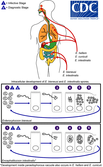
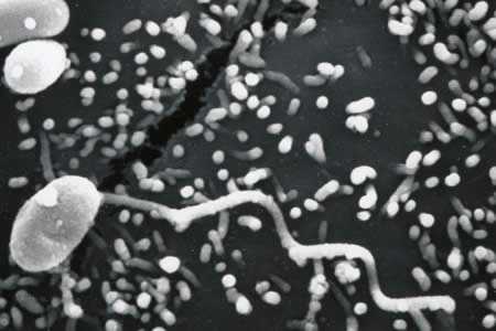
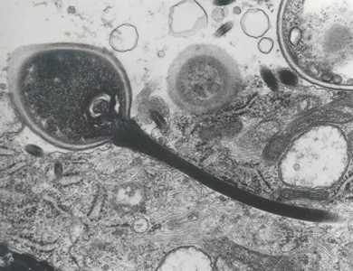
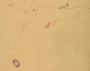
The infective form of microsporidia is the resistant spore and it can survive for a long time in the environment  . The spore extrudes its polar tubule and infects the host cell
  . The spore injects the infective sporoplasm into the eukaryotic host cell through the polar tubule
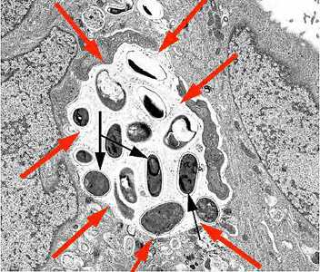
  . Inside the cell, the sporoplasm undergoes extensive multiplication either by merogony (binary fission) or schizogony (multiple fission)
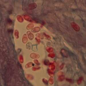
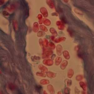
  . This development can occur either in direct contact with the host cell cytoplasm (e.g., E. bieneusi) or inside a vacuole termed parasitophorous vacuole (e.g., E. intestinalis). Either free in the cytoplasm or inside a parasitophorous vacuole, microsporidia develop by sporogony to mature spores
  . During sporogony, a thick wall is formed around the spore, which provides resistance to adverse environmental conditions. When the spores increase in number and completely fill the host cell cytoplasm, the cell membrane is disrupted and releases the spores to the surroundings
  . These free mature spores can infect new cells thus continuing the cycle.
Geographic Distribution
Microsporidia are being increasingly recognized as opportunistic infectious agents worldwide.
Clinical Presentation
Human microsporidiosis represents an important and rapidly emerging opportunistic disease, occurring mainly, but not exclusively, in severely immunocompromised patients with AIDS. Additionally, cases of microsporidiosis in immunocompromised persons not infected with HIV as well as in immunocompetent persons also have been reported. The clinical manifestations of microsporidiosis are very diverse, varying according to the causal species with diarrhea being the most common.
| Microsporidian species | Clinical manifestation |
|---|---|
| Anncaliia algerae | Keratoconjunctivitis, skin and deep muscle infection |
| Enterocytozoon bieneusi* | Diarrhea, acalculous cholecystitis |
| Encephalitozoon cuniculi and Encephalitozoon hellem | Keratoconjunctivitis, infection of respiratory and genitourinary tract, disseminated infection |
| Encephalitozoon intestinalis (syn. Septata intestinalis) | Infection of the GI tract causing diarrhea, and dissemination to ocular, genitourinary and respiratory tracts |
| Microsporidium (M. ceylonensis and M. africanum) | Infection of the cornea |
| Nosema sp. (N. ocularum), Anncaliia connori | Ocular infection |
| Pleistophora sp. | Muscular infection |
| Trachipleistophora anthropophthera | Disseminated infection |
| Trachipleistophora hominis | Muscular infection, stromal keratitis, (probably disseminated infection) |
| Tubulinosema acridophagus | Disseminated infection |
| Vittaforma corneae (syn. Nosema corneum) | Ocular infection, urinary tract infection |
*Two reports of E. bieneusi in respiratory samples have also been published, one in 1992 and the other in 1997.

DPDx is an education resource designed for health professionals and laboratory scientists. For an overview including prevention and control visit www.cdc.gov/parasites/.
- Page last reviewed: May 3, 2016
- Page last updated: May 3, 2016
- Content source:
- Global Health – Division of Parasitic Diseases and Malaria
- Notice: Linking to a non-federal site does not constitute an endorsement by HHS, CDC or any of its employees of the sponsors or the information and products presented on the site.
- Maintained By: