Case #269 - February, 2010

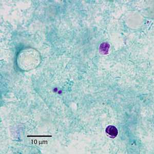
Figure A

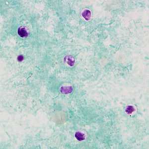
Figure B

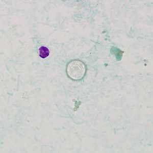
Figure C
Case Answer
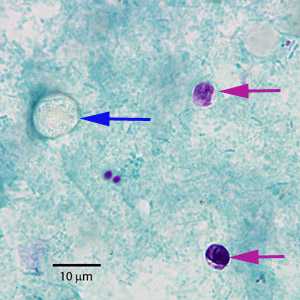
This case represented a mixed infection with Cyclospora sp. and Cryptosporidium sp. Diagnostic morphologic features shown in the figures were:
- a size range for the unstained objects which was consistent with Cyclospora sp. (blue arrow, Figure A).
- unstained oocysts with a wrinkled appearance in the modified acid-fast stain, another characteristic of Cyclospora sp. All of the oocysts shown in this case were unstained, which is not uncommon.
- a size range for the stained objects which was consistent with Cryptosporidium sp. (red arrow, Figure A).
- the presence of sporozoites in some of the Cryptosporidium oocysts.
Morphology can only identify Cryptosporidium and Cyclospora to the genus level. Molecular testing (PCR) of appropriate specimens should be performed to identify the parasites to the species level.

Figure A
More on: Cryptosporidiosis: Cyclosporiasis
Images presented in the monthly case studies are from specimens submitted for diagnosis or archiving. On rare occasions, clinical histories given may be partly fictitious.
DPDx is an education resource designed for health professionals and laboratory scientists. For an overview including prevention and control visit www.cdc.gov/parasites/.
- Page last reviewed: August 24, 2016
- Page last updated: August 24, 2016
- Content source:
- Global Health – Division of Parasitic Diseases and Malaria
- Notice: Linking to a non-federal site does not constitute an endorsement by HHS, CDC or any of its employees of the sponsors or the information and products presented on the site.
- Maintained By:


 ShareCompartir