Case #287 - November, 2010
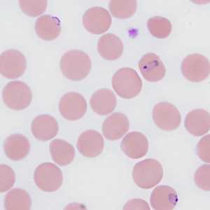
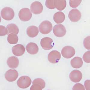
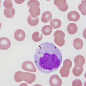
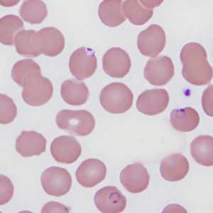
A 29-year-old pregnant woman from Ethiopia presented to a hospital with nausea, vomiting, abdominal pain and episodic fever. The symptoms started within a day after arriving in the United States, and had persisted for five days prior to hospitalization. Given her country of origin, blood specimens were collected in EDTA for malaria screening. She reported having been treated for malaria approximately one year prior (medications unknown). Images were captured from a Wright-stained thin blood smear at 1000x magnification and sent via email to the DPDx Team for diagnostic assistance. Figures A-D show four of the images sent to DPDx. What is your diagnosis? Based on what criteria?

Figure A

Figure B

Figure C

Figure D
Case Answer
This was a case of malaria caused by Plasmodium falciparum. Diagnostic morphologic features included small, delicate rings with small, chromatin dots in normal-sized (not enlarged) infected red blood cells. Appliqué forms were also present in Figures A, B, and D. While Figure C did not show any parasites, the white blood cell shown did show evidence of ingested malarial pigment.
More on: Malaria
Images presented in the monthly case studies are from specimens submitted for diagnosis or archiving. On rare occasions, clinical histories given may be partly fictitious.
DPDx is an education resource designed for health professionals and laboratory scientists. For an overview including prevention and control visit www.cdc.gov/parasites/.
- Page last reviewed: August 24, 2016
- Page last updated: August 24, 2016
- Content source:
- Global Health – Division of Parasitic Diseases and Malaria
- Notice: Linking to a non-federal site does not constitute an endorsement by HHS, CDC or any of its employees of the sponsors or the information and products presented on the site.
- Maintained By:


 ShareCompartir