
Case #340 - February, 2013
The DPDx Team received a pair of proglottids from a state health lab for cestode confirmation and identification. The specimens were submitted in 70% ethanol and measured on average 12.0 mm long by 3.0 mm wide. The proglottids were reportedly found in the feces of a 43-year-old woman with no documented international travel. Figures A and B show one of the proglottids. Figures C and D show the same proglottid after soaking in lactophenol for several hours. What is your diagnosis? Based on what criteria?
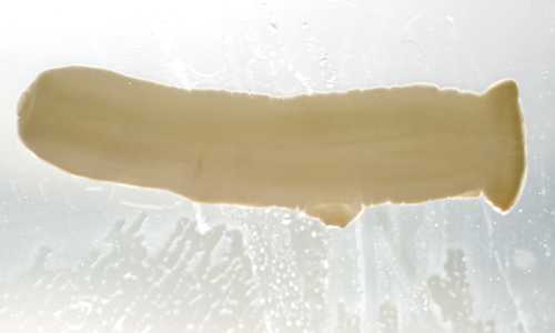
Figure A
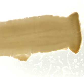
Figure B
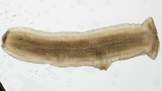
Figure C
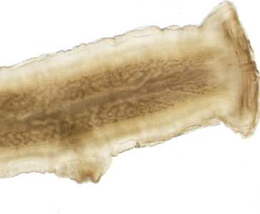
Figure D
Case Answer
This was a case of taeniasis caused by the beef tapeworm, Taenia saginata. Diagnostic morphologic features included:
- a single, prominent lateral genital pore (GP, Figure D).
- more than 13 primary uterine branches on either side of the central uterine stem (Figures C and D).
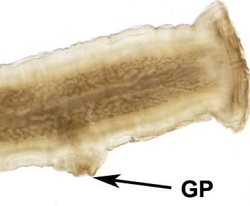
Figure D
More on: Taeniasis
Images presented in the monthly case studies are from specimens submitted for diagnosis or archiving. On rare occasions, clinical histories given may be partly fictitious.
DPDx is an education resource designed for health professionals and laboratory scientists. For an overview including prevention and control visit www.cdc.gov/parasites/.
- Page last reviewed: August 24, 2016
- Page last updated: August 24, 2016
- Content source:
- Global Health – Division of Parasitic Diseases and Malaria
- Notice: Linking to a non-federal site does not constitute an endorsement by HHS, CDC or any of its employees of the sponsors or the information and products presented on the site.
- Maintained By:


 ShareCompartir