Case #441 - April, 2017
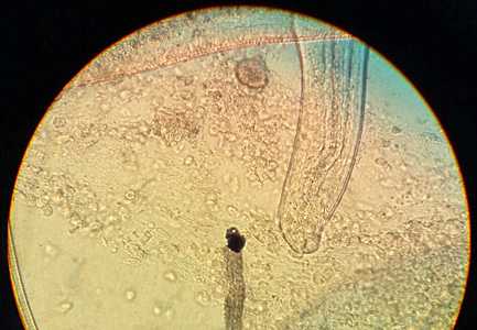
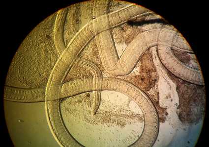
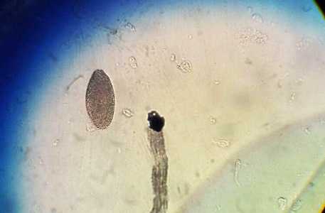
A patient, who lives in a rural region in South America, presented with bloody diarrhea and remembers having eaten undercooked pork. The attending physician submitted images to the DPDx Team of a roundworm he observed during a colonoscopy. Based on the inner structures observed which appeared to be stichocytes, Trichinella spp. was initially suspected as the causative agent. However, an ELISA test for trichinellosis was negative. Figures A-C show what was observed from the colonoscopy. What is your diagnosis? Based on what criteria?

Figure A

Figure B

Figure C
Case Answer
This was a case of trichuriasis casued by Trichuris trichuris (also referred to as whipworm) In addition to location in the patient being consistent with T. trichuria (colon) vs. Trichinella sp. (usual location is the small intestine). Other morphologic features shown include
- A very long esophagus with stichosomes (Figure B).
- A simple mouth with no lips (Figure A).
- An immature egg (Figure C) that although does not demonstrate the typical polor plugs of T. trichuria, does rule-out Trichinella spp.
More on: Trichuris trichiura
This case and images were kindly provided by The University of Buenos Aires, University Hospital, Parasitology Division, Buenos Aires, Argentina.
Images presented in the monthly case studies are from specimens submitted for diagnosis or archiving. On rare occasions, clinical histories given may be partly fictitious.
DPDx is an education resource designed for health professionals and laboratory scientists. For an overview including prevention and control visit www.cdc.gov/parasites/.
- Page last reviewed: May 11, 2017
- Page last updated: May 17, 2017
- Content source:
- Global Health – Division of Parasitic Diseases and Malaria
- Notice: Linking to a non-federal site does not constitute an endorsement by HHS, CDC or any of its employees of the sponsors or the information and products presented on the site.
- Maintained By:


 ShareCompartir