
Case #445 - June, 2017
Several people developed gastrointestinal symptoms of watery diarrhea, nausea with vomiting, and low-grade fever approximately 1 week after attending a catered event. Stool specimens were collected for laboratory testing which included a formalin-ethyl acetate concentration with brightfield wet mount examination. Figures A–D show what was observed at 400x magnification in all of the specimens. The objects of interest shown ranged in size from 8 to 10 micrometers What is your diagnosis? What other test(s), if any, would you recommend?
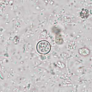
Figure A
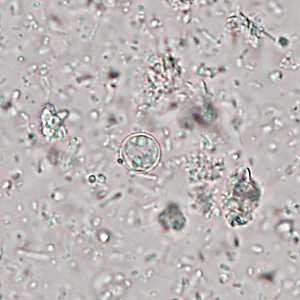
Figure B
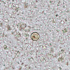
Figure C
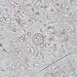
Figure D
Case Answer
These were cases of cyclosporiasis caused by Cyclospora cayetanensis.
Diagnostic features included round, refractile, non-sporulated oocysts within the size range for Cyclospora (Figures A, B, and D).
Although accurate identification of Cyclospora using bright-field microscopy is possible on unstained wet mount preparations, we recommend using UV microscopy to enhance sensitivity and specificity. Figures A, B, and C show the same objects that were presented using bright field microscopy and UV microscopy. Note how it is evident that the object in Figure C is not Cyclospora based on the fact that the entire object fluoresces and not just the oocytst wall as shown in true oocysts in Figures A and B.
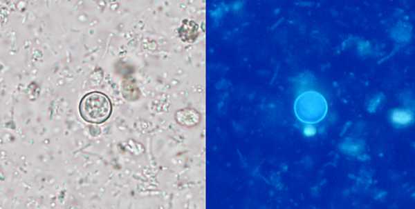
Figure A
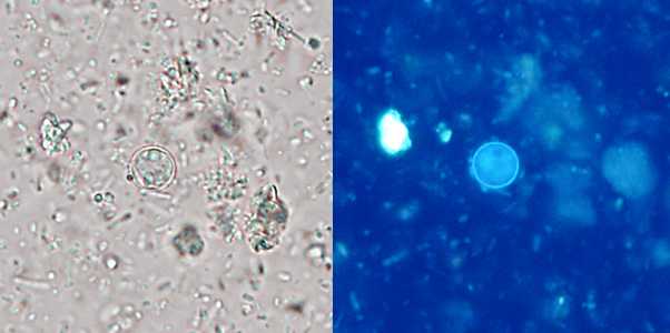
Figure B
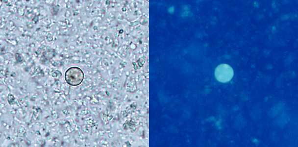
Figure C
More on Cyclosporiasis
Images presented in the monthly case studies are from specimens submitted for diagnosis or archiving. On rare occasions, clinical histories given may be partly fictitious.
DPDx is an education resource designed for health professionals and laboratory scientists. For an overview including prevention and control visit www.cdc.gov/parasites/.
- Page last reviewed: July 19, 2017
- Page last updated: July 19, 2017
- Content source:
- Global Health – Division of Parasitic Diseases and Malaria
- Notice: Linking to a non-federal site does not constitute an endorsement by HHS, CDC or any of its employees of the sponsors or the information and products presented on the site.
- Maintained By:


 ShareCompartir