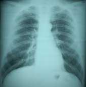
CHEST RADIOGRAPHY

Radiographic Classification: Epidemiologic Research
There is a long and distinguished history of using the International Labour Office (ILO) Classification System to derive classifications for input to epidemiologic studies and related activities (i.e., health surveillance and health hazard investigations). Not only have the results of these studies proven the validity of the ILO Classification System and associated methodology but they have led to hundreds of scientific papers on disease prevalence and its relationship with exposure, time, place, job, and industry. Many findings have been applied to risk analysis, and used for workplace compliance standard setting (e.g., coal mine dust).
The Role of Classification of Chest Radiographs in Epidemiologic Research
Pneumoconioses form an important class of occupational respiratory disease, and are a major public health concern. There has been a continuing need not only to estimate disease extent and severity over time and place, but to correlate measures of abnormality with indices of exposure so that exposure-response models can be developed. These findings have provided input to risk analysis, leading to recommendations for occupational exposure levels in the workplace. Surveillance programs enable disease tracking over time and place, indicating whether prevention measures are effective. Hazard investigations provide data on potential risk to workers.
The validity of ILO Classification System has been repeatedly demonstrated in many settings and industries. For example, classifications of radiographs of coal miners show clear correlations with dust exposure, lung dust burden, lung pathology, and mortality (Attfield 1992, Ruckley 1984, Miller 1985). Elsewhere, for example, classifications of radiographs of patients with asbestos-related lung disease were shown to be correlated with lung function (Cotes 1988).
Special Considerations for Classification of Chest Radiographs in Epidemiologic Research
A chest radiograph classification is one of many other health outcome measurements that have been applied to epidemiologic research. As with any other measurement in epidemiology, a fundamental objective is to ensure that it meets certain data quality standards: that is, it is necessary to pay attention to accuracy and precision considerations. Traditionally, researchers in research and surveillance have been aware of the need for quality assurance when obtaining classifications for epidemiologic research, and experienced researchers are familiar with the many ways to ensure good data quality. These include procedures for selecting readers, training and assessing readers in pilot studies, simultaneous quality control, use of multiple readers, and use of unbiased summary scoring methods. A useful summary of criteria to consider for epidemiologic purposes is given by Mulloy et al. (1993).
Factors Relevant to Classification of Chest Radiographs in Epidemiologic Research
1. ILO Classification System
Use of the ILO Classification System will help maintain consistency with accepted standards of abnormality, ensure uniform standards within a program, and ensure comparability with other data.
2. Remuneration
Remuneration that is based on individual classification outcomes or on the overall level of reported abnormality has the obvious potential to cause bias.
3. Reader selection
Readers should have demonstrated skills (e.g., B Readers) and experience. Prior reader selection procedures can be applied, in which preliminary classification exercises are undertaken to assess reader levels, permit training, and eliminate outliers as necessary.
4. Number of readings and summary classifications
The ILO (ILO 2002) recommends a minimum of two classifications, but states that preferably more be obtained. If two classifications are obtained, a third reader can be employed to classify the radiographs where the first two readers disagree, thereby enabling the median of three classifications to be computed for all radiographs. Where resources permit, researchers may prefer to obtain three (or more) independent classifications of all radiographs, because of the higher scientific flexibility and quality that ensues.
Independent classifications from multiple readers are typically combined into a single summary classification. Summarization methods that are unbiased, (i.e., represent the middle of the distribution of classifications), such as use of the median, are preferable. As an alternative to summary classifications, some studies have undertaken analyses by each reader’s classifications and averaged the resulting statistics. Use of reader panels in which a summary classification is derived through discussion of the radiograph is not recommended as it has been shown to favor those readers who dominate or are most senior.
5. Blinding
When classified radiographs are used for epidemiologic purposes, it is essential to be aware that knowledge of ancillary details specific to individuals can introduce bias into results. This includes medical or exposure information and other readers’ interpretations. (ILO 2002) To avoid the effects of any temporal changes in classification practices, radiographs should be randomly allocated to readers.
6. Quality assurance
Quality assurance procedures designed to reduce inter-reader variation before the start of a study and to monitor and correct problems during the course of the chest radiograph classification will help optimize the reliability of a study’s findings. Simultaneous quality assurance, done by placing unidentified quality control (“calibration”) radiographs with a previously established array of parenchymal and/or pleural findings within the set of unknown radiographs being evaluated, provides the most realistic assessment of how readers classify unknown radiographs. Providing feedback comparing the reader’s classification of these radiographs to the previously-established classifications has been used to maintain and improve reader performance (Sheers 1978). A National Institutes of Health-sponsored workshop suggested including chest radiographs of unexposed workers in epidemiologic studies for purposes of control (Weill 1975).
In extended reading exercises, in order to avoid the effects of any temporal changes in classification practices across all readers, the readers should classify batches of radiographs in random order.
7. Notification
Whenever possible and especially when individual medical findings are pertinent to maintaining and protecting that individual’s health, it is ethically necessary to inform individuals of findings from their individual chest radiograph. Prior unblinded readings may be necessary to provide workers with the best information on their health. However, the information obtained from the research classifications, including the individual and summary classifications, should also be conveyed to the examinee. There is ethical justification in notifying member of occupational cohorts and their employers of results of scientific investigations in which they participate. To further disease identification and to promote prevention, reporting of diagnosed or suspected cases of pneumoconiosis to state public health organizations is required in some states.
References
Attfield MD, Morring K. An investigation into the relationship between coal workers’ pneumoconiosis and dust exposure in U.S. coal miners. Am Ind Hyg Assoc J 1992; 53:486-92.
Ruckley VA, Fernie JM, Chapman JS, et al. Comparison of radiographic appearance with associated pathology and lung dust content in a group of coalworkers. Br J Ind Med 1984; 41:459-67.
Miller BG, Jacobsen M. Dust exposure, pneumoconiosis, and mortality of coal miners. Br J Ind Med 1985; 42:723-33.
Cotes JE, King B. Relationship of lung function to radiographic classification (ILO) in patients with asbestos related lung disease. Thorax 1988; 43(10):777-83.
Mulloy KB, Coultas DB, Samet JM. Use of chest radiographs in epidemiological investigations of pneumoconioses. Br J Ind Med 1993; 50(3):273-5.
Fay JWJ, Rae S. The Pneumoconiosis Field Research of the National Coal Board. Ann Occup Hyg 1959; 1:149-61.
Hurley JF, Burns J, Copland L, et al. Coalworkers’ simple pneumoconiosis and exposure to dust at 10 British coalmines. Br J Ind Med 1982; 39:120-7.
Sheers G, Rossiter CE, Gilson JC, et al. UK naval dockyards asbestos study: radiological methods in the surveillance of workers exposed to asbestos. Br J Ind Med 1978; 35:195-203.
Weill H, Jones R. The chest roentgenogram as an epidemiologic tool. Report of a workshop. Arch Environ Health 1975; 30:435-9.
- Page last reviewed: May 24, 2011
- Page last updated: November 17, 2011
- Content source:
- National Institute for Occupational Safety and Health Respiratory Health Division


 ShareCompartir