Bankart lesion
| Bankart lesion | |
|---|---|
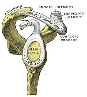 | |
| The glenoid labrum, labeled glenoid ligament, is damaged in a Bankart lesion. Side view demonstrating the articular surface of the right scapula is shown. | |
| Specialty | Orthopedics |
| Types | Variations: Anterior labroligamentous periostous sleeve avulsion lesion (ALPSA), reverse Bankart lesion[1] |
| Treatment | Medical imaging; X-ray, CT scan, MRI scan[2] |
A Bankart lesion is a type of shoulder injury that occurs following a dislocated shoulder.[1] The anterior (inferior) glenoid labrum of the shoulder detaches.[2] When this happens, a pocket at the front of the glenoid forms that allows the humeral head to come out of the shoulder joint.[2] There is not always a fracture.[1] Other variations include anterior labroligamentous periostous sleeve avulsion lesion (ALPSA), and reverse Bankart lesion.[1]
Diagnosis is by medical imaging; X-ray, CT scan, MRI scan.[2] It is an indication for surgery and often accompanied by a Hill-Sachs lesion, damage to the posterior humeral head.[3]
The Bankart lesion is named after English orthopedic surgeon Arthur Sydney Blundell Bankart (1879–1951).[4]
Diagnosis
The diagnosis is usually initially made by a combination of physical exam and medical imaging, where the latter may be projectional radiography (in cases of bony Bankart) and/or MRI of the shoulder. The presence of intra-articular contrast allows for better evaluation of the glenoid labrum.[5] Type V SLAP tears extends into the Bankart defect.[6]
Treatment
Arthroscopic repair of Bankart injuries have good success rates, though nearly one-third of patients require further surgery for continued instability after the initial procedure in a study of young adults, with higher re-operation rates in those less than 20 years of age.[7] Options for repair include an arthroscopic technique or a more invasive open Latarjet procedure,[8] with the open technique tending to have a lower incidence of recurrent dislocation, but also a reduced range of motion following surgery.[9]
Gallery
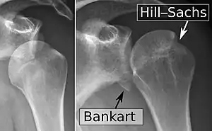 X-ray at left shows anterior dislocation in a young man after trying to get up from his bed. X-ray at right shows same shoulder after reduction and internal rotation, revealing both a bony Bankart lesion and a Hill-Sachs lesion.
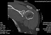 CT scan showing a bony Bankart lesion at the antero-inferior glenoid
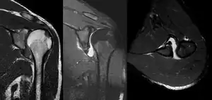 MRI of the shoulder after an anterior dislocation showing a Hill-Sachs lesion and labral Bankart lesion
 Bankart lesion seen at arthroscopy
 Radiograph showing a bony Bankart lesion with stationary fragment at the inferior glenoid
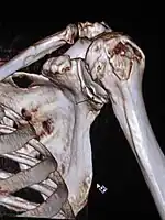 3-D CT reconstruction of a bankart lesion which occurred post anterior shoulder dislocation. This subject's humerus remains mildly superiorly subluxated. Fracture marked by a black arrow.
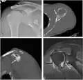 Bony Bankart
References
- 1 2 3 4 Major, Nancy M.; Anderson, Mark W. (2020). "10. Shoulder". Musculoskeletal MRI. Philadelphia: Elsevier. pp. 218–219. ISBN 978-0-323-415606. Archived from the original on 2022-09-20. Retrieved 2022-09-12.
- 1 2 3 4 Martel, José; Bueno, Angel (2008). "Fractures with names". In Pope, Thomas; Bloem, Hans L.; Beltran, Javier; Morrison, William B.; John, David (eds.). Musculoskeletal Imaging (2nd ed.). Philadelphia: Elsevier. p. 1232.e2. ISBN 978-1-4557-0813-0. Archived from the original on 2022-09-20. Retrieved 2022-09-11.
- ↑ Porcellini, Giuseppe; Campi, Fabrizio; Paladini, Paolo (2002). "Arthroscopic approach to acute bony Bankart lesion". Arthroscopy: The Journal of Arthroscopic and Related Surgery. 18 (7): 764–769. doi:10.1053/jars.2002.35266. ISSN 0749-8063. PMID 12209435.
- ↑ Hunter, Tim B.; Peltier, Leonard F.; Lund, Pamela J. (1 May 2000). "Radiologic History Exhibit". RadioGraphics. pp. 819–836. doi:10.1148/radiographics.20.3.g00ma20819. Archived from the original on 16 September 2022. Retrieved 16 September 2022.
- ↑ Jana, M; Srivastava, DN; Sharma, R; Gamanagatti, S; Nag, H; Mittal, R; Upadhyay, AD (April 2011). "Spectrum of magnetic resonance imaging findings in clinical glenohumeral instability". The Indian Journal of Radiology & Imaging. 21 (2): 98–106. doi:10.4103/0971-3026.82284. PMC 3137866. PMID 21799591.
- ↑ Chang, D; Mohana-Borges, A; Borso, M; Chung, CB (October 2008). "SLAP lesions: anatomy, clinical presentation, MR imaging diagnosis and characterization". European Journal of Radiology. 68 (1): 72–87. doi:10.1016/j.ejrad.2008.02.026. PMID 18499376.
- ↑ Flinkkilä, T; Knape, R; Sirniö, K; Ohtonen, P; Leppilahti, J (16 March 2017). "Long-term results of arthroscopic Bankart repair: Minimum 10 years of follow-up". Knee Surgery, Sports Traumatology, Arthroscopy. 26 (1): 94–99. doi:10.1007/s00167-017-4504-z. PMID 28303281. S2CID 6692528. Archived from the original on 21 January 2021. Retrieved 29 November 2021.
- ↑ Zimmermann, SM; Scheyerer, MJ; Farshad, M; Catanzaro, S; Rahm, S; Gerber, C (7 December 2016). "Long-Term Restoration of Anterior Shoulder Stability: A Retrospective Analysis of Arthroscopic Bankart Repair Versus Open Latarjet Procedure" (PDF). The Journal of Bone and Joint Surgery. American Volume. 98 (23): 1954–1961. doi:10.2106/jbjs.15.01398. PMID 27926676. Archived from the original on 7 February 2020. Retrieved 29 November 2021.
- ↑ Wang, L; Liu, Y; Su, X; Liu, S (8 October 2015). "A Meta-Analysis of Arthroscopic versus Open Repair for Treatment of Bankart Lesions in the Shoulder". Medical Science Monitor. 21: 3028–35. doi:10.12659/msm.894346. PMC 4603609. PMID 26446430.