Fibulin
| Anaphylotoxin-like domain | |||||||||||
|---|---|---|---|---|---|---|---|---|---|---|---|
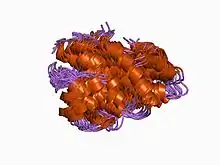
 Structure of porcine C5adesArg.[1] | |||||||||||
| Identifiers | |||||||||||
| Symbol | ANATO | ||||||||||
| Pfam | PF01821 | ||||||||||
| InterPro | IPR000020 | ||||||||||
| SMART | ANATO | ||||||||||
| PROSITE | PDOC00906 | ||||||||||
| SCOP2 | 1c5a / SCOPe / SUPFAM | ||||||||||
| CDD | cd00017 | ||||||||||
| |||||||||||
Fibulin (FY-beau-lin) (now known as Fibulin-1 FBLN1) is the prototypic member of a multigene family, currently with seven members. Fibulin-1 is a calcium-binding glycoprotein. In vertebrates, fibulin-1 is found in blood and extracellular matrices. In the extracellular matrix, fibulin-1 associates with basement membranes and elastic fibers. The association with these matrix structures is mediated by its ability to interact with numerous extracellular matrix constituents including fibronectin, proteoglycans, laminins and tropoelastin. In blood, fibulin-1 binds to fibrinogen and incorporates into clots.
Fibulins are secreted glycoproteins that become incorporated into a fibrillar extracellular matrix when expressed by cultured cells or added exogenously to cell monolayers.[2][3] The five known members of the family share an elongated structure and many calcium-binding sites, owing to the presence of tandem arrays of epidermal growth factor-like domains. They have overlapping binding sites for several basement-membrane proteins, tropoelastin, fibrillin, fibronectin and proteoglycans, and they participate in diverse supramolecular structures. The amino-terminal domain I of fibulin consists of three anaphylatoxin-like (AT) modules, each approximately 40 residues long and containing four or six cysteines. The structure of an AT module was determined for the complement-derived anaphylatoxin C3a, and was found to be a compact alpha-helical fold that is stabilized by three disulphide bridges in the pattern Cys14, Cys25 and Cys36 (where Cys is cysteine). The bulk of the remaining portion of the fibulin molecule is a series of nine EGF-like repeats.[4]
Genes
- FBLN1, FBLN2, FBLN3, FBLN4, FBLN5, FBLN7 and HMCN1 is also known as "fibulin-6".[5]
References
- ↑ Williamson MP, Madison VS (March 1990). "Three-dimensional structure of porcine C5adesArg from 1H nuclear magnetic resonance data". Biochemistry. 29 (12): 2895–905. doi:10.1021/bi00464a002. PMID 2337573.
- ↑ Burgess WH, Argraves WS, Tran H, Dickerson K (1990). "Fibulin is an extracellular matrix and plasma glycoprotein with repeated domain structure". J. Cell Biol. 111 (6): 3155–3164. doi:10.1083/jcb.111.6.3155. PMC 2116371. PMID 2269669.
- ↑ Timpl R, Sasaki T, Kostka G, Chu ML (2003). "Fibulins: a versatile family of extracellular matrix proteins". Nat. Rev. Mol. Cell Biol. 4 (6): 479–489. doi:10.1038/nrm1130. PMID 12778127. S2CID 8442153.
- ↑ Timpl R, Sasaki T, Chu ML, Pan TC, Zhang RZ, Fassler R (1993). "Structure and expression of fibulin-2, a novel extracellular matrix protein with multiple EGF-like repeats and consensus motifs for calcium binding". J. Cell Biol. 123 (5): 1269–1277. doi:10.1083/jcb.123.5.1269. PMC 2119879. PMID 8245130.
- ↑ Fisher SA, Rivera A, Fritsche LG, et al. (April 2007). "Case-control genetic association study of fibulin-6 (FBLN6 or HMCN1) variants in age-related macular degeneration (AMD)". Hum. Mutat. 28 (4): 406–13. doi:10.1002/humu.20464. PMID 17216616.
External links
- Fibulin at the US National Library of Medicine Medical Subject Headings (MeSH)
- Argraves WS, Tran H, Burgess WH, Dickerson K (1990). "Fibulin is an extracellular matrix and plasma glycoprotein with repeated domain structure". J. Cell Biol. 111 (6 Pt 2): 3155–64. doi:10.1083/jcb.111.6.3155. PMC 2116371. PMID 2269669.