Inferior suprarenal artery
| Inferior suprarenal artery | |
|---|---|
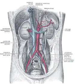
 Posterior abdominal wall, after removal of the peritoneum, showing kidneys, suprarenal capsules, and great vessels | |
| Details | |
| Source | Renal artery |
| Vein | Suprarenal veins |
| Supplies | Adrenal gland |
| Identifiers | |
| Latin | Arteria suprarenalis inferior |
| TA98 | A12.2.12.077 |
| TA2 | 4271 |
| FMA | 69264 |
| Anatomical terminology | |
The inferior suprarenal artery is a paired artery that supplies the adrenal gland. It usually originates at the trunk of the renal artery before its terminal division, but with many common variations. It supplies the adrenal gland parenchyma, the ureter, and the surrounding cellular tissue and muscles.
Structure
The inferior suprarenal artery usually originates at the trunk of the renal artery.[1][2] This is usually on its superior surface before its terminal division.[1] It enters the parenchyma of the adrenal gland.[1]
Variations
Variations in the interior suprarenal artery are common.[1][3] It usually originates from the renal artery before its final divisions, but may also originate as a final division or after the final divisions.[1] More rarely, it may originate directly from the aorta.[1] It may give off a small branch to the kidney.[1]
There may be two or three inferior suprarenal arteries in some people.[1] Its diameter changes significantly with age.[4]
Function
The inferior suprarenal artery supplies the adrenal gland (suprarenal gland).[1] They also supply the ureter and some surrounding tissue and skeletal muscle.
Clinical significance
The inferior suprarenal artery may be affected by an aneurysm.[5] It may be assessed using Doppler ultrasound.[6]
History
The inferior suprarenal artery may also be known as the inferior adrenal artery.[6]
See also
References
![]() This article incorporates text in the public domain from page 610 of the 20th edition of Gray's Anatomy (1918)
This article incorporates text in the public domain from page 610 of the 20th edition of Gray's Anatomy (1918)
- 1 2 3 4 5 6 7 8 9 Bordei, P.; D. Şt Antohe; E. Şapte; D. Iliescu (2003). "Morphological aspects of the inferior suprarenal artery". Surgical and Radiologic Anatomy. 25 (3–4): 247–251. doi:10.1007/s00276-003-0132-z. PMID 14504822. S2CID 9934169.
- ↑ Chiva, Luis M.; Magrina, Javier (2018). "2 - Abdominal and Pelvic Anatomy". Principles of Gynecologic Oncology Surgery. Elsevier. pp. 3–49. doi:10.1016/B978-0-323-42878-1.00002-X. ISBN 978-0-323-42878-1.
- ↑ Deepthinath, R.; B. Satheesha Nayak; R.B. Mehta; Seetharama Bhat; Vincent Rodrigues; Vijaya Paul Samuel; V. Venkataramana; A.M. Prasad (2006). "Multiple variations in the paired arteries of the abdominal aorta". Clinical Anatomy. 19 (6): 566–568. doi:10.1002/ca.20207. PMID 16283657. S2CID 43834737.
- ↑ Toni, R.; S. Mosca; L. Favero; S. Ricci; R. Roversi; G. Toni; P. Vezzadini (1988). "Clinical anatomy of the suprarenal arteries: a quantitative approach by aortography". Surgical and Radiologic Anatomy. 10 (4): 297–302. doi:10.1007/BF02107902. PMID 3145571. S2CID 24843738.
- ↑ Manners, James; Singh, Rajinder; Page, Andrew; Adamson, Andrew; McLean, Duncan (December 2010). "Radiological treatment of a spontaneously ruptured inferior adrenal artery aneurysm". Nature Reviews Urology. 7 (12): 694–698. doi:10.1038/nrurol.2010.184. ISSN 1759-4820.
- 1 2 Fujita, Yasuyuki; Satoh, Shoji; Nakano, Hitoo (2001-10-01). "Doppler velocimetry in the adrenal artery in human fetuses". Early Human Development. 65 (1): 47–55. doi:10.1016/S0378-3782(01)00196-7. ISSN 0378-3782.
External links
- Anatomy figure: 40:02-09 at Human Anatomy Online, SUNY Downstate Medical Center – "The suprarental glands. Blood supply to the suprarenal glands."
- Anatomy photo:40:04-0103 at the SUNY Downstate Medical Center – "Posterior Abdominal Wall: Blood Supply to the Suprarenal Glands"
- Photo of model at Waynesburg College circulation/leftinferiorsuprarenalartery