Left colic artery
| Left colic artery | |
|---|---|
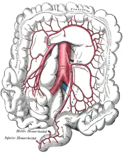
 The inferior mesenteric artery and its branches. (Left colic visible at center right.) | |
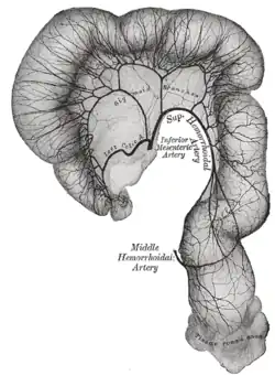
 Sigmoid colon and rectum, showing distribution of branches of inferior mesenteric artery and their anastomoses. (Left colic visible at center left.) | |
| Details | |
| Source | inferior mesenteric |
| Vein | left colic vein |
| Supplies | descending colon |
| Identifiers | |
| Latin | arteria colica sinistra |
| TA98 | A12.2.12.071 |
| TA2 | 4292 |
| FMA | 14826 |
| Anatomical terminology | |
The left colic artery is a branch of the inferior mesenteric artery.
Structure
It runs to the left behind the peritoneum and in front of the psoas major muscle. After a short, but variable, course, it divides into an ascending and a descending branch. The stem of the artery or its branches cross the left ureter and left internal spermatic vessels.
The ascending branch crosses in front of the left kidney and ends, between the two layers of the transverse mesocolon, by anastomosing with the middle colic artery; the descending branch anastomoses with the highest sigmoid artery.
From the arches formed by these anastomoses branches are distributed to the descending colon and the left part of the transverse colon.
Clinical significance
The left colic artery may be ligated during abdominal surgery to remove colorectal cancer.[1] This may have poorer outcomes than preserving the artery.[1]
Additional images
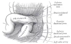
 Superior and inferior duodenal fossæ.
 Duodenojejunal fossa.
References
![]() This article incorporates text in the public domain from page 610 of the 20th edition of Gray's Anatomy (1918)
This article incorporates text in the public domain from page 610 of the 20th edition of Gray's Anatomy (1918)
- 1 2 Fan, Yu-Chen; Ning, Fei-Long; Zhang, Chun-Dong; Dai, Dong-Qiu (April 2018). "Preservation versus non-preservation of left colic artery in sigmoid and rectal cancer surgery: A meta-analysis". International Journal of Surgery (London, England). 52: 269–277. doi:10.1016/j.ijsu.2018.02.054. ISSN 1743-9159. PMID 29501795.
External links
- Lotti M. Anatomy in relation to left colectomy
- sup&infmesentericart at The Anatomy Lesson by Wesley Norman (Georgetown University)
- Anatomy photo:39:05-0105 at the SUNY Downstate Medical Center - "Intestines and Pancreas: Branches of the Inferior Mesenteric Artery"
- Anatomy image:8585 at the SUNY Downstate Medical Center
- Anatomy image:8658 at the SUNY Downstate Medical Center