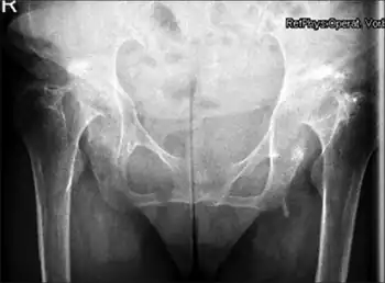
Protrusio acetabuli
| Protrusio acetabuli | |
|---|---|
 | |
| Radiographic criteria for protrusio acetabuli are an abnormally positioned acetabular line, a center-edge angle of Wiberg of >40°, and the crossing of teardrop by ilio-ischial line | |
| Specialty | Orthopedics |
Protrusio acetabuli is an uncommon defect of the acetabulum, the socket that receives the femoral head to make the hip joint. The hip bone of the pelvic bone/girdle is composed of three bones, the ilium, the ischium and the pubis. In protrusio deformity, there is medial displacement of the femoral head in that the medial aspect of the femoral cortex is medial to the ilioischial line. The socket is too deep and may protrude into the pelvis.[1]
Signs and symptoms
Protrusio acetabuli may be asymptomatic. Limitation of joint range of movement is the earliest sign, along with pain.
Diagnosis
Classification
Protrusio acetabuli is divided into two types, primary and secondary.
- Primary protrusio acetabuli are characterized by progressive protrusio in middle aged women, and may be associated with osteoarthritis. They may be familial.
- Secondary protrusio acetabuli's causes include femoral head prosthesis, cup arthroplasty, septic arthritis, central fracture dislocation, or total hip replacement surgery
Protrusio acetabuli may also be thought of as unilateral or bilateral.
- Unilateral protrusio acetabuli may be caused by tuberculous arthritis, trauma, or fibrous dysplasia.
- Bilateral protrusio acetabuli may be caused by rheumatoid arthritis, Paget's disease, osteomalacia,[2] Marfan syndrome,[3] and ankylosing spondylitis.
Treatment
Arthroscopic surgery (or open joint surgery) is an effective treatment. Joint replacement surgery may be necessary in the case of severe pain or substantial joint restriction.
Prognosis
The protrusio may progress until the femoral neck impinges against the pelvis.
References
- ↑ Van De Velde S, Fillman R, Yandow S (2006). "The aetiology of protrusio acetabuli. Literature review from 1824 to 2006". Acta Orthop Belg. 72 (5): 524–9. PMID 17152413.
- ↑ Dahnert's Radiology
- ↑ Van de Velde S, Fillman R, Yandow S (2006). "Protrusio acetabuli in Marfan syndrome. History, diagnosis, and treatment". J Bone Joint Surg Am. 88 (3): 639–46. doi:10.2106/JBJS.E.00567. PMID 16510833.
{{cite journal}}: CS1 maint: url-status (link)
External links
| Classification |
|---|
- McBride MT, Muldoon MP, Santore RF, Trousdale RT, Wenger DR (2001). "Protrusio acetabuli: diagnosis and treatment". J Am Acad Orthop Surg. 9 (2): 79–88. doi:10.5435/00124635-200103000-00002. PMID 11281632. Archived from the original on 2023-02-09. Retrieved 2022-01-22.