Rosenthal fiber

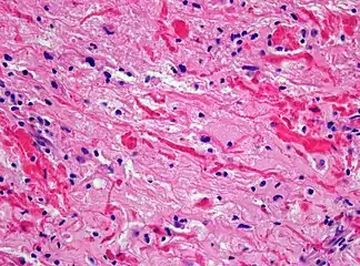
H&E staining showing these elongated eosinophilic structures in a case of pilocytic astrocytoma. Magnification 400x
A Rosenthal fiber is a thick, elongated, worm-like or "corkscrew" eosinophilic (pink) bundle that is found on staining of brain tissue in the presence of long-standing gliosis, occasional tumors, and some metabolic disorders.
Associated conditions
Its presence is associated with either pilocytic astrocytoma[1] (more common) or Alexander's disease (a rare leukodystrophy). They are also seen in the context of fucosidosis.
Pilocytic astrocytoma is the most common primitive tumor found in pediatrics.
Composition
The fibers are found in astrocytic processes and are thought to be clumped intermediate filament proteins, primarily glial fibrillary acidic protein.[2] Other reported constituents include alphaB crystallin, heat shock protein 27, protein beta-1), ubiquitin, vimentin, plectin, c-Jun, the 20 S proteasome, and synemin.[3]
References
- ↑ Wippold FJ, Perry A, Lennerz J (May 2006). "Neuropathology for the neuroradiologist: Rosenthal fibers". AJNR Am J Neuroradiol. 27 (5): 958–61. PMID 16687524.
- ↑ Tanaka KF, Ochi N, Hayashi T, Ikeda E, Ikenaka K (October 2006). "Fluoro-Jade: new fluorescent marker of Rosenthal fibers". Neurosci. Lett. 407 (2): 127–30. doi:10.1016/j.neulet.2006.08.014. PMID 16949206.
- ↑ Heaven, MR; Flint, D; Randall, SM; et al. (July 1, 2016). "Composition of Rosenthal Fibers, the Protein Aggregate Hallmark of Alexander Disease". Journal of Proteome Research. 15 (7): 2265–82. doi:10.1021/acs.jproteome.6b00316. PMC 5036859. PMID 27193225.
External links
- Neuropathology Mini-Course. Chapter 9 - Tumors of the Nervous System
- Doctor's Doctor - Brain and Spinal Cord
- Isolation of a major protein component of Rosenthal fibers