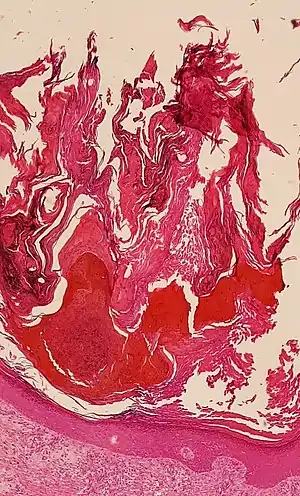
Hyperkeratosis
Hyperkeratosis is thickening of the stratum corneum (the outermost layer of the epidermis, or skin), often associated with the presence of an abnormal quantity of keratin,[1] and also usually accompanied by an increase in the granular layer. As the corneum layer normally varies greatly in thickness in different sites, some experience is needed to assess minor degrees of hyperkeratosis.
| Hyperkeratosis | |
|---|---|
 | |
| Micrograph showing prominent hyperkeratosis in skin without atypia. H&E stain. | |
| Pronunciation |
|
| Specialty | Dermatology |
It can be caused by vitamin A deficiency or chronic exposure to arsenic.
Hyperkeratosis can also be caused by B-Raf inhibitor drugs such as Vemurafenib and Dabrafenib.[2]
It can be treated with urea-containing creams, which dissolve the intercellular matrix of the cells of the stratum corneum, promoting desquamation of scaly skin, eventually resulting in softening of hyperkeratotic areas.[3]
Types
Follicular
Follicular hyperkeratosis, also known as keratosis pilaris (KP), is a skin condition characterized by excessive development of keratin in hair follicles, resulting in rough, cone-shaped, elevated papules. The openings are often closed with a white plug of encrusted sebum. When called phrynoderma the condition is associated with nutritional deficiency or malnourishment.
This condition has been shown in several small-scale studies to respond well to supplementation with vitamins and fats rich in essential fatty acids. Deficiencies of vitamin E,[4] vitamin A, and B-complex vitamins have been implicated in causing the condition.[5] Follicular hyperkeratosis is also a symptom in inherited collagen-related diseases of Ehlers-Danlos syndromes and Bethlem myopathy.
By other specific site
Hereditary
- Epidermolytic hyperkeratosis (also known as "Bullous congenital ichthyosiform erythroderma,"[7] "Bullous ichthyosiform erythroderma,"[8]: 482 or "bullous congenital ichthyosiform erythroderma of Brocq"[9]) is a rare skin disease in the ichthyosis family affecting around 1 in 250,000 people. It involves the clumping of keratin filaments.[6]: 562 [10]
- Multiple minute digitate hyperkeratosis, a rare cutaneous condition, with about half of cases being familial
- Focal acral hyperkeratosis (also known as "Acrokeratoelastoidosis lichenoides,") is a late-onset keratoderma, inherited as an autosomal dominant condition, characterized by oval or polygonal crateriform papules developing along the border of the hands, feet, and wrists.[8]: 509
- Keratosis pilaris appears similar to gooseflesh, is usually asymptomatic and may be treated by moisturizing the skin.[11]
In mucous membranes
The term hyperkeratosis is often used in connection with lesions of the mucous membranes, such as leukoplakia. Because of the differences between mucous membranes and the skin (e.g. keratinizing mucosa does not have a stratum lucidum and non keratinizing mucosa does not have this layer or normally a stratum corneum or a stratum granulosum), sometimes specialized texts give slightly different definitions of hyperkeratosis in the context of mucosae. Examples are "an excessive formation of keratin (e.g., as seen in leukoplakia)"[12] and "an increase in the thickness of the keratin layer of the epithelium, or the presence of such a layer in a site where none would normally be expected."[13]
Etymology and pronunciation
The word hyperkeratosis (/ˌhaɪpərˌkɛrəˈtoʊsɪs/) is based on the Ancient Greek morphemes hyper- + kerato- + -osis, meaning 'the condition of too much keratin'.
Hyperkeratosis in dogs
Nasodigitic hyperkeratosis in dogs may be idiopathic, secondary to an underlying disease, or due to congenital abnormalities in the normal anatomy of the nose and fingertips.
In the case of congenital anatomical abnormalities, contact between the affected area and rubbing surfaces is impaired. It is roughly the same with finger pads - in animals with an anatomical abnormality part of the pad is not in contact with rubbing surfaces and excessive keratin deposition is formed. The idiopathic form of nasodigitic hyperkeratosis in dogs develops from unknown causes and is more common in older animals (senile form).[14][15] Of all dog breeds, Labradors, Golden Retrievers, Cocker Spaniels, Irish Terriers, Bordeaux Dogs are the most prone to hyperkeratosis.[16]
Therapy
Since the deposition of excess keratin cannot be stopped, therapy is aimed at softening and removing it. For moderate to severe cases, the affected areas should be hydrated (moisturised) with warm water or compresses for 5-10 minutes. Softening preparations are then applied once a day until the excess keratin is removed.
In dogs with severe hyperkeratosis and a significant excess of keratin, it is removed with scissors or a blade. After proper instructions, pet owners are able to perform this procedure at home and it may be the only method of correction.[17]
See also
References
- Kumar, Vinay; Fausto, Nelso; Abbas, Abul (2004) Robbins & Cotran Pathologic Basis of Disease (7th ed.). Saunders. Page 1230. ISBN 0-7216-0187-1.
- Niezgoda, Anna; Niezgoda, Piotr; Czajkowski, Rafal (2015) Novel Approaches to Treatment of Advanced Melanoma: A Review of Targeted Therapy and Immunotherapy BioMed Research International
- drugs.com > Urea Cream (Prescribing Information) Revised: 04/2010 by Stratus Pharmaceuticals
- Nadiger, HA (1980). "Role of vitamin E in the aetiology of phrynoderma (follicular hyperkeratosis) and its interrelationship with B-complex vitamins". British Journal of Nutrition. 44 (3): 211–4. doi:10.1079/bjn19800033. PMID 7437404.
- "Hyperkeratosis". Dorland's Medical Dictionary for Health Consumers. 2007.
- James, William D.; Berger, Timothy G.; Elston, Dirk M.; et al. (2006). "Clinical diagnosis by laboratory methods". Andrews' diseases of the skin: clinical dermatology (10th ed.). Saunders Elsevier. ISBN 0-7216-2921-0.
- Rapini, Ronald P.; Bolognia, Jean L.; Jorizzo, Joseph L. (2007). Dermatology: 2-Volume Set. St. Louis: Mosby. ISBN 978-1-4160-2999-1.
- Freedberg, et al. (2003). Fitzpatrick's Dermatology in General Medicine. (6th ed.). McGraw-Hill. ISBN 0-07-138076-0.
- synd/1036 at Who Named It?
- Cheng J, Syder AJ, Yu QC, Letai A, Paller AS, Fuchs E (September 1992). "The genetic basis of epidermolytic hyperkeratosis: a disorder of differentiation-specific epidermal keratin genes". Cell. 70 (5): 811–9. doi:10.1016/0092-8674(92)90314-3. PMID 1381287. S2CID 31906051.
- Hwang, Schwartz (Sep 2008). "Keratosis pilaris: a common follicular hyperkeratosis". Cutis. 82 (3): 177–80. PMID 18856156.
- Mosby's Dental Dictionary
- Tyldesley WR, Field A, Longman L (2003). Tyldesley's Oral medicine (5th ed.). Oxford: Oxford University Press. ISBN 0192631470.
- "Muller and Kirk's Small Animal Dermatology. Saunders, St. Luis, MO". scholar.google.com. Retrieved December 16, 2021.
- "Skin diseases of the dog and cat: clinical and histopathologic diagnosis". scholar.google.com. Retrieved December 16, 2021.
- "What Does it Mean When a Dog's Nose is Dry? When to Worry". boneandyarn.com. 8 August 2020. Retrieved December 16, 2021.
- "Hyperkeratosis In Dogs: Does Your Dog Have Hairy Feet?". caninejournal.com. 7 January 2016. Retrieved December 16, 2021.