Slime mold
Slime mold or slime mould is an informal name given to a polyphyletic assemblage of unrelated eukaryotic organisms in the Stramenopiles, Rhizaria, Discoba, Amoebozoa and Holomycota. Most are microscopic; those in the Myxogastria form larger plasmodial slime molds visible to the naked eye. The slime mold life cycle includes a free-living single-celled stage and the formation of spores. Spores are often produced in macroscopic multicellular or multinucleate fruiting bodies which may be formed through aggregation or fusion; aggregation is driven by chemical signals called acrasins. Slime molds contribute to the decomposition of dead vegetation; some are parasitic.

Slime molds have a variety of behaviors otherwise seen in animals with brains. Species such as Physarum polycephalum have been used to simulate traffic networks.
Evolution
Taxonomic history

Two groups, the Myxomycetes or plasmodial slime molds, and the Acrasieae or cellular slime molds, were described by the French botanist Philippe van Tieghem in 1880.[2] In 1859 and 1887, the German mycologist Heinrich Anton de Bary placed both groups in a new class, Mycetozoa, with a section of "Doubtful Mycetozoa" for Plasmodiophora (now part of Phytomyxea) and Labyrinthula. He stated that these were neither plants nor fungi.[2] In 1868, the German biologist Ernst Haeckel placed the Mycetozoa in a kingdom he named Protista.[2] In 1885, the British zoologist Ray Lankester grouped the Mycetozoa alongside the Proteomyxa as part of the Gymnomyxa in the phylum Protozoa.[2]
In 1956, the American biologist Herbert Copeland placed the Mycetozoa (the myxomycetes and plasmodiophorids) and the Sarkodina (the labyrinthulids and the cellular slime molds) in a phylum called Protoplasta, which he placed alongside the fungi and the algae in a new kingdom, Protoctista.[2][3]
In 1969, the taxonomist R. H. Whittaker stated that the "slime molds stick out like a sore thumb" from the rest of the Fungi, to which they were at that time attached, and agreed to Lindsay S. Olive's reassignment of the Gymnomycota to the Protista.[4] Whittaker placed three phyla, namely the Myxomycota, Acrasiomycota, and Labyrinthulomycota in a subkingdom Gymnomycota within the Fungi.[2]
Phylogeny
Slime molds have little or no fossil history, as might be expected given that they are small and soft-bodied.[5] The grouping is polyphyletic, consisting of multiple clades (emphasised in the phylogenetic tree) widely scattered across the Eukaryotes:[6][7]
| Eukaryotes |
| ||||||||||||||||||||||||||||||||||||||||||||||||||||||||||||||||||||||||
Diversity
Macroscopic, plasmodial slime molds: Myxogastria
The Myxogastria or plasmodial slime molds are the only macroscopic scale slime molds; they gave the group its informal name, since for part of their life cycle they are slimy to the touch.[8] Most are smaller than a few centimetres, but some species may reach sizes up to several square metres and masses up to 20 kilograms.[9][10][11] They consist of a large cell with thousands of nuclei within a single membrane without walls, forming a syncytium.[12]
_with_Ant.jpg.webp)
_Rostaf_1014107_(cropped).jpg.webp)

_M.L._Farr%252C_1976_(Reticularia_lycoperdon)_(cropped).JPG.webp) Enteridium lycoperdon, mid-sporangial phase
Enteridium lycoperdon, mid-sporangial phase
Cellular slime molds: Dictyosteliida
The Dictyosteliida or cellular slime molds do not form huge coenocytes like the Myxogastria; their amoebae remain individual for most of their lives as individual unicellular protists, feeding on microorganisms. However, when food is depleted and they are ready to form sporangia, they release signal molecules into their environment, by which they find each other and create swarms. These amoebae then join up into a tiny multicellular slug-like coordinated creature, which crawls to an open lit place and grows into a fruiting body, a sorocarp. Some of the amoebae become spores to begin the next generation, but others sacrifice themselves to become a dead stalk, lifting the spores up into the air.[13][14]
Protosteliida
The protosteloids, a polyphyletic group, have characters intermediate between the previous two groups, but they are much smaller, the fruiting bodies only forming one to a few spores.[15]
Non-amoebozoan slime molds
Among the non-amoebozoan slime molds are the Acrasids, which have sluglike amoebae have eruptive pseudopodia.[16] The Phytomyxea are obligate parasites, with hosts among the plants, diatoms, oomycetes, and brown algae. They cause plant diseases like cabbage club root and powdery scab.[17] The Labyrinthulomycetes are marine slime nets, forming labyrinthine networks of tubes in which amoeba without pseudopods can travel.[18] The Fonticulida are cellular slime molds that form a fruiting body in a "volcano" shape.[19]
Life cycle
Plasmodial slime molds

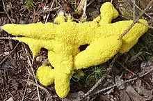
Plasmodial slime molds begin life as amoeba-like cells. These unicellular amoebae are commonly haploid and feed on bacteria. These amoebae can mate if they encounter the correct mating type and form zygotes that then grow into plasmodia. These contain many nuclei without cell membranes between them, and can grow to meters in size. The species Fuligo septica is often seen as a slimy yellow network in and on rotting logs. The amoebae and the plasmodia engulf microorganisms.[20] The plasmodium grows into an interconnected network of protoplasmic strands.[21] Within each protoplasmic strand, the cytoplasmic contents rapidly stream, periodically reversing direction. The streaming protoplasm within a plasmodial strand can reach speeds of up to 1.35 mm per second, the fastest for any microorganism.[22]
Slime molds are isogamous, which means that their gametes (reproductive cells) are all the same size, unlike the eggs and sperms of animals.[23] Physarum polycephalum has three reproductive genes – matA, matB, and matC. The first two of these have thirteen variants. MatC, however, only has three variants. Each reproductively mature slime mold is diploid, meaning that it contains two copies of each of the three reproductive genes.[24] When P. polycephalum is ready to make its reproductive cells, it grows a bulbous extension of its body to contain them.[25] Each cell has a random combination of the genes that the slime mold contains within its genome. Therefore, it can create cells of up to eight different gene types. Once these cells are released, they are independent and tasked with finding another cell it is able to fuse with. Other P. polycephalum may contain different combinations of the matA, matB, and matC genes, allowing over 500 possible variations. It is advantageous for organisms with this type of reproductive cell to have many mating types because the likelihood of the cells finding a partner is greatly increased, and the risk of inbreeding is drastically reduced.[24]
Cellular slime molds
The cellular slime molds exist as single-celled organisms while food is plentiful. When food is in short supply, many of these single-celled organisms congregate and start moving as a single body. In this state they are sensitive to airborne chemicals and can detect food sources. They readily change the shape and function of parts, and may form stalks that produce fruiting bodies, releasing countless spores, light enough to be carried on the wind or on passing animals.[13] The cellular slime mold Dictyostelium discoideum has many different mating types. When this organism has entered the stage of reproduction, it releases an attractant, called acrasin. Acrasin is made up of cyclic adenosine monophosphate, or cyclic AMP. Cyclic AMP is crucial in passing hormone signals between reproductive cells.[26] When it comes time for the cells to fuse, Dictyostelium discoideum has mating types of its own that dictate which cells are compatible with each other. There are at least eleven mating types; macrocysts form after cell contact between compatible mating types.[27]
Chemical signals

The chemicals that aggregate cellular slime molds are small molecules called acrasins. The first acrasin to be discovered was cyclic adenosine monophosphate (cyclic AMP), in Dictyostelium discoideum. During the aggregation phase of their life cycle, Dictyostelium discoideum amoebae communicate with each other by traveling waves of cyclic AMP.[28][29][30] There is an amplification of cyclic AMP when they aggregate.[31] Pre-stalk cells move toward cyclic AMP, but pre-spore cells ignore the signal.[32] The acrasin for Polysphondylium violaceum was purified in 1983; it is a dipeptide that has been named glorin. Its major components are the amino acids glutamic acid and ornithine. An amino group (NH3) and a carboxyl group (COOH) of the glutamic acid are blocked respectively by a propionyl group and an ethyl ester. An amino group on the ornithine molecule is blocked by a lactam ring.[33]
Behavior
Similarity to neural systems
Slime molds share some similarities with neural systems in animals.[34] The membranes of both slime molds and neural cells contains receptor sites, which alter electrical properties of the membrane when it is bound.[35] Therefore, some studies on the early evolution of animal neural systems are inspired by slime molds.[36][37][38] When a slime mold mass or mound is physically separated, the cells find their way back to re-unite. Studies on Physarum polycephalum have even shown an ability to learn and predict periodic unfavorable conditions in laboratory experiments.[39] John Tyler Bonner, a professor of ecology known for his studies of slime molds, argues that they are "no more than a bag of amoebae encased in a thin slime sheath, yet they manage to have various behaviors that are equal to those of animals who possess muscles and nerves with ganglia – that is, simple brains."[40]
Traffic system inspirations

Atsushi Tero and colleagues grew Physarum in a flat wet dish, placing the mold in a central position representing Tokyo, and oat flakes surrounding it corresponding to the locations of other major cities in the Greater Tokyo Area. As Physarum avoids bright light, light was used to simulate mountains, water and other obstacles in the dish. The mold first densely filled the space with plasmodia, and then thinned the network to focus on efficiently connected branches. The network closely resembled Tokyo's rail system.[41][42] P. polycephalum was used in experimental laboratory approximations of motorway networks of 14 geographical areas: Australia, Africa, Belgium, Brazil, Canada, China, Germany, Iberia, Italy, Malaysia, Mexico, the Netherlands, UK and US.[43][44][45] The filamentary structure of P. polycephalum forming a network to food sources is similar to the large scale galaxy filament structure of the universe. This observation has led astronomers to use simulations based on the behaviour of slime molds to inform their search for dark matter.[46][47]
See also
- Swarming motility – rapid and coordinated translocation of a bacterial population across solid or semi-solid surfaces
- Water mold – Fungus-like eukaryotic microorganism
References
- Lister, Arthur; Lister, Gulielma (1911). A monograph of the Mycetozoa : a descriptive catalogue of the species in the Herbarium of the British Museum. London: Printed by order of the Trustees of the British Museum. doi:10.5962/bhl.title.21191.
- Olive, Lindsay S.; Stoianovitch, Carmen (technical assistance) (1975). The Mycetozoans. Academic Press. pp. 1–7. ISBN 978-0-1252-6250-7.
- Copeland, H. F. (1956). The Classification of Lower Organisms. Palo Alto, California: Pacific Books.
- Whittaker, R. H. (16 May 1969). "Response: Reassignment of Gymnomycota". Science. American Association for the Advancement of Science (AAAS). 164 (3881): 857–857. doi:10.1126/science.164.3881.857.b. ISSN 0036-8075.
- "Introduction to the 'Slime Molds'". University of California Museum of Paleontology.
- Vallverdú, Jordi; et al. (2018). "Slime mould: the fundamental mechanisms of biological cognition". BioSystems (165): 57–70.
- Baldauf, S.L.; Doolittle, W.F. (October 1997). "Origin and Evolution of the Slime Molds (Mycetozoa)". PNAS. 94 (22): 12007–120012. Bibcode:1997PNAS...9412007B. doi:10.1073/pnas.94.22.12007. PMC 23686. PMID 9342353.
- Adamatzky, Andrew (2016). Advances in Physarum Machines: Sensing and Computing with Slime Mould. Springer. p. 4. ISBN 978-3-319-26662-6.
- Ing, B. (1999). The myxomycetes of Britain and Ireland: an identification handbook. Slough, England: Richmond Publishing. p. 4. ISBN 978-0-85546-251-2.
- Nannenga-Bremekamp, N.E. (1974). De Nederlandse Myxomyceten. Zuthpen: Koninklijke Nederlandse Natuurhistorische Vereniging. ISBN 978-90-03-93130-6.
- Zhulidov, Daniel A.; Robarts, Richard D.; Zhulidov, Alexander V.; Zhulidova, Olga V.; Markelov, Danila A.; Rusanov, Viktor A.; Headley, John V. (2002). "Zinc accumulation by the slime mold Fuligo septica (L.) Wiggers in the former Soviet Union and North Korea". Journal of Environmental Quality. 31 (3): 1038–1042. doi:10.2134/jeq2002.1038. PMID 12026071.
- Ples, Marek (2023-11-11). "Lab Snapshots by Marek Ples; Microbiology - The biology on a different level". weirdscience.eu. Retrieved 2023-07-02.
- Jacobson, R. (April 5, 2012). "Slime Molds: No Brains, No Feet, No Problem". PBS Newshour.
- Kin, K.; Schaap, P. (March 2021). "Evolution of Multicellular Complexity in The Dictyostelid Social Amoebas". Genes. 12 (4): 487. doi:10.3390/genes12040487. PMC 8067170. PMID 33801615.
- Fiore-Donno, Anna Maria; Nikolaev, Sergey I.; Nelson, Michaela; Pawlowski, Jan; Cavalier-Smith, Thomas; Baldauf, Sandra L. (January 2010). "Deep Phylogeny and Evolution of Slime Moulds (Mycetozoa)". Protist. 161 (1): 55–70. doi:10.1016/j.protis.2009.05.002. PMID 19656720.
- Brown, Matthew W.; Silberman, Jeffrey D.; Spiegel, Frederick W. (2012). "A contemporary evaluation of the acrasids (Acrasidae, Heterolobosea, Excavata)". European Journal of Protistology. Elsevier BV. 48 (2): 103–123. doi:10.1016/j.ejop.2011.10.001. ISSN 0932-4739.
- Neuhauser, Sigrid; Kirchmair, Martin; Bulman, Simon; Bass, David (2014). "Cross-kingdom host shifts of phytomyxid parasites". BMC Evolutionary Biology. 14 (1): 33. doi:10.1186/1471-2148-14-33. PMC 4016497. PMID 24559266.
- Tsui, Clement K. M.; Marshall, Wyth; Yokoyama, Rinka; Honda, Daiske; Lippmeier, J Casey; Craven, Kelly D.; Peterson, Paul D.; Berbee, Mary L. (January 2009). "Labyrinthulomycetes phylogeny and its implications for the evolutionary loss of chloroplasts and gain of ectoplasmic gliding". Molecular Phylogenetics and Evolution. 50 (1): 129–40. doi:10.1016/j.ympev.2008.09.027. PMID 18977305.
- Deasey, Mary C.; Olive, Lindsay S. (July 1981). "Role of Golgi Apparatus in Sorogenesis by the Cellular Slime Mold Fonticula alba". Science. 213 (4507): 561–563. Bibcode:1981Sci...213..561D. doi:10.1126/science.213.4507.561. PMID 17794844.
- Ling, H. (2012). "Myxomycetes: Overlooked Native Plants". The Native Plant Society of New Jersey. Archived from the original on 9 June 2015. Retrieved 29 May 2018.
- Chimileski, Scott; Kolter, Roberto. "Life at the Edge of Sight". www.hup.harvard.edu. Harvard University Press. Retrieved 2018-01-26.
- Alexopoulos, C.J. (1962). Introductory Mycology (Second ed.). New York, N.Y.: John Wiley and Sons. p. 78.
- Moskvitch, Katia (9 July 2018). "Slime Molds Remember – but Do They Learn?". Quanta Magazine. Retrieved 2019-11-02.
- Judson, Olivia (2002). Dr. Tatiana's Sex Advice To All Creation. New York: Henry Holt and Company. pp. 187–193. ISBN 978-0-8050-6332-5.
- Renner, B. (2006). "Slime Mold Reproduction". BioWeb. University of Wisconsin System. Retrieved 2019-11-02.
- Bonner, J.T. (2009). The Social Amoebae: The Biology of Cellular Slime Molds. Princeton University Press. ISBN 978-0-691-13939-5. JSTOR j.ctt7s6qz.
- Erdos, Gregory W.; Raper, Kenneth B.; Vogen, Linda K. (June 1973). "Mating Types and Macrocyst Formation in Dictyostelium discoideum". Proceedings of the National Academy of Sciences of the United States of America. 70 (6): 1828–1830. Bibcode:1973PNAS...70.1828E. doi:10.1073/pnas.70.6.1828. PMC 433606. PMID 16592095.
- Nestle, Marion; Sussman, Maurice (August 1972). "The effect of cyclic AMP on morphogernesis and enzyme accumulation in Dictyostelium discoideum". Developmental Biology. 28 (4): 545–554. doi:10.1016/0012-1606(72)90002-4. PMID 4340352.
- Levine, Herbert; Reynolds, William (May 1991). "Streaming instability of aggregating slime mold amoebae". Physical Review Letters. 66 (18): 2400–2403. Bibcode:1991PhRvL..66.2400L. doi:10.1103/PhysRevLett.66.2400. PMID 10043475.
- Tyson, John J.; Alexander, Kevin A.; Manoranjan, V. S.; Murray, J.D. (1989-01-01). "Spiral waves of cyclic amp in a model of slime mold aggregation". Physica D: Nonlinear Phenomena. 34 (1): 193–207. Bibcode:1989PhyD...34..193T. doi:10.1016/0167-2789(89)90234-0. ISSN 0167-2789.
- Roos, W.; Nanjundiah, V.; Malchow, D.; Gerisch, G. (May 1975). "Amplification of cyclic-AMP signals in aggregating cells of Dictyostelium discoideum". FEBS Letters. 53 (2): 139–142. doi:10.1016/0014-5793(75)80005-6. PMID 166875. S2CID 29448450.
- Fujimori, Taihei; Nakajima, Akihiko; Shimada, Nao; Sawai, Satoshi (March 2019). "Tissue self-organization based on collective cell migration by contact activation of locomotion and chemotaxis". Proceedings of the National Academy of Sciences of the United States of America. 116 (10): 4291–4296. Bibcode:2019PNAS..116.4291F. doi:10.1073/pnas.1815063116. PMC 6410881. PMID 30782791.
- Bonner, John Tyler (1983). "Chemical Signals of Social Amoebae". Scientific American. 248 (4): 114–121. ISSN 0036-8733. JSTOR 24968880.
- Carr, William E. S. (1989). "Chemical Signaling Systems in Lower Organisms: A Prelude to the Evolution of Chemical Communication in the Nervous System". In Anderson, Peter A.V. (ed.). Evolution of the First Nervous Systems. Boston, MA: Springer. pp. 81–94. doi:10.1007/978-1-4899-0921-3_6. ISBN 978-1-4899-0921-3.
- Carr, William E. S.; Gleeson, Richard A.; Trapido-Rosenthal, Henry G. (June 1990). "The role of perireceptor events in chemosensory processes". Trends in Neurosciences. 13 (6): 212–215. doi:10.1016/0166-2236(90)90162-4. PMID 1694326. S2CID 46452914.
- Lindsey, J.; Lasker, R. (1974). "Chemical Signals in the Sea: Marine Allelochemics and Evolution". Fishery Bulletin. 72 (1): 1–11.
- Lenhoff, H M; Heagy, W (April 1977). "Aquatic invertebrates: model systems for study of receptor activation and evolution of receptor proteins". Annual Review of Pharmacology and Toxicology. 17 (1): 243–258. doi:10.1146/annurev.pa.17.040177.001331. PMID 17353.
- Janssens, P.M.; Van Haastert, P.J. (December 1987). "Molecular basis of transmembrane signal transduction in Dictyostelium discoideum". Microbiological Reviews. 51 (4): 396–418. doi:10.1128/mr.51.4.396-418.1987. PMC 373123. PMID 2893972.
- Saigusa, Tetsu; Tero, Atsushi; Nakagaki, Toshiyuki; Kuramoto, Yoshiki (January 2008). "Amoebae anticipate periodic events". Physical Review Letters. 100 (1): 018101. Bibcode:2008PhRvL.100a8101S. doi:10.1103/PhysRevLett.100.018101. hdl:2115/33004. PMID 18232821.
- Barone, Jennifer (December 8, 2008). "#71: Slime Molds Show Surprising Degree of Intelligence". Discover Magazine.
- MacPherson, Kitta (January 21, 2010). "The 'sultan of slime': Biologist continues to be fascinated by organisms after nearly 70 years of study". Princeton University.
- Tero, A.; Takagi, S.; Saigusa, T.; et al. (January 2010). "Rules for biologically inspired adaptive network design" (PDF). Science. 327 (5964): 439–442. Bibcode:2010Sci...327..439T. doi:10.1126/science.1177894. PMID 20093467. S2CID 5001773. Archived from the original (PDF) on 2013-04-21.
- Yong, Ed (January 21, 2010). "Slime mould attacks simulates Tokyo rail network". ScienceBlogs.
- Christiansen B (25 January 2010). "Slime Mold Network Engineering". Technovelgy.
- Marks, P. (6 January 2010). "Designing highways the slime mould way". New Scientist.
- Adamatzky, Andrew; Akl, S.; Alonso-Sanz, R.; et al. (2013). "Are motorways rational from slime mould's point of view?". International Journal of Parallel, Emergent and Distributed Systems. 28 (3): 230–248. arXiv:1203.2851. doi:10.1080/17445760.2012.685884. S2CID 15534238.
- Parr, D. (18 February 2014). "Cities in motion: how slime mould can redraw our rail and road maps". The Guardian.
- "Slime Mold Simulations Used to Map Dark Matter". NASA. 10 March 2020.
- Wenz, J. (12 March 2020). "Slime mold helps astronomers map dark matter". Astronomy magazine.