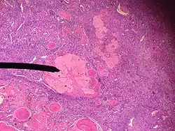
Corpus albicans
The corpus albicans (Latin for "whitening body"; also known as atretic corpus luteum, corpus candicans, or simply as albicans) is the regressed form of the corpus luteum. As the corpus luteum is being broken down by macrophages, fibroblasts lay down type I collagen, forming the corpus albicans. This process is called "luteolysis". The remains of the corpus albicans may persist as a scar on the surface of the ovary.
| Corpus albicans | |
|---|---|
 Human corpus albicans | |
| Details | |
| Identifiers | |
| Latin | Corpus albicans |
| TA98 | A09.1.01.016 |
| TA2 | 3485 |
| FMA | 18620 |
| Anatomical terminology | |
Background
During the first few hours after expulsion of the ovum from the follicle, the remaining granulosa and theca interna cells change rapidly into lutein cells. They enlarge in diameter two or more times and become filled with lipid inclusions that give them a yellowish appearance.
This process is called luteinization, and the total mass of cells together is called the corpus luteum. A well-developed vascular supply also grows into the corpus luteum.
The granulosa cells in the corpus luteum develop extensive intracellular smooth endoplasmic reticula that form large amounts of the female sex hormones progesterone and estrogen (more progesterone than estrogen during the luteal phase). The theca cells form mainly the androgens androstenedione and testosterone rather than female sex hormones. However, most of these hormones are also converted by the enzyme aromatase in the granulosa cells into estrogens, the female hormones.
The corpus luteum normally grows to about 1.5 centimeters in diameter, reaching this stage of development 7 to 8 days after ovulation. Then it begins to involute and eventually loses its secretory function and its yellowish, lipid characteristic about 12 days after ovulation, becoming the corpus albicans;[1] during the ensuing few weeks, this is replaced by connective tissue and over months is absorbed.[2]
References
- Marieb, Elaine (2013). Anatomy & physiology. Benjamin-Cummings. p. 915. ISBN 9780321887603.
- Guyton and Hall Textbook of Medical Physiology (12th Ed) 2011, page number: 991
- "corpus albicans", Stedman's Online Medical Dictionary at Lippincott Williams and Wilkins
- Hiatt, James L.; Gartner, Leslie P. (2001). Color textbook of histology. Philadelphia: W.B. Saunders. ISBN 0-7216-8806-3.
External links
- Histology image: 97_03 at the University of Oklahoma Health Sciences Center
- Histology image: 18104loa – Histology Learning System at Boston University
- Histology at KUMC female-female09