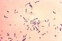
Corynebacterium diphtheriae
Corynebacterium diphtheriae[lower-alpha 1] is the pathogenic bacterium that causes diphtheria.[2] It is also known as the Klebs–Löffler bacillus, because it was discovered in 1884 by German bacteriologists Edwin Klebs (1834–1912) and Friedrich Löffler (1852–1915).[3] The bacteria are harmless unless they are infected by a bacteriophage that carries a gene that gives rise to a toxin. This toxin causes the disease.[4]
| Corynebacterium diphtheriae | |
|---|---|
 | |
| Scientific classification | |
| Domain: | Bacteria |
| Phylum: | Actinomycetota |
| Class: | Actinomycetia |
| Order: | Mycobacteriales |
| Family: | Corynebacteriaceae |
| Genus: | Corynebacterium |
| Species: | C. diphtheriae |
| Binomial name | |
| Corynebacterium diphtheriae (Kruse 1886) Lehmann and Neumann 1896 (Approved Lists 1980)[1] | |
Classification
Four subspecies are recognized: C. d. mitis, C. d. intermedius, C. d. gravis, and C. d. belfanti. The four subspecies differ slightly in their colonial morphology and biochemical properties, such as the ability to metabolize certain nutrients, but all may be toxigenic (and therefore cause diphtheria) or not toxigenic. C. diphtheriae produces diphtheria toxin which alters protein function in the host by inactivating the elongation factor EF-2. This causes pharyngitis and 'pseudomembrane' in the throat. The strains which are toxigenic are ones which have been infected with a bacteriophage.[5][6]
The diphtheria toxin gene is encoded by the bacteriophage found in toxigenic strains, integrated into the bacterial chromosome.[7] To accurately identify C. diphtheriae, a Gram stain is performed to show Gram-positive, highly pleomorphic organisms with no particular arrangement. Special stains like Albert's stain and Ponder's stain are used to demonstrate the metachromatic granules formed in the polar regions. The granules are called polar granules, Babes Ernst granules or volutin granules. An enrichment medium, such as Löffler's medium, is used to preferentially grow C. diphtheriae. After that, a differential plate known as tellurite agar, allows all Corynebacteria (including C. diphtheriae) to reduce tellurite to metallic tellurium. The tellurite reduction is colorimetrically indicated by brown colonies for most Cornyebacterium species or by a black halo around the C. diphtheriae colonies.
A low concentration of iron is required in the medium for toxin production. At high iron concentrations, iron molecules bind to an aporepressor on the beta bacteriophage, which carries the Tox gene. When bound to iron, the aporepressor shuts down toxin production.[8] Elek's test for toxigenicity is used to determine whether the organism is able to produce the diphtheria toxin.[9]
Pathogenicity

Corynebacterium diphtheriae is the bacterium that causes the disease called diphtheria. Bacteriophages introduce a gene into the bacterial cells that makes a strain toxigenic. The strains that are not infected with these viruses are harmless.[4] C. diphtheriae is a rod-shaped, Gram-positive, nonspore-forming, and nonmotile bacterium.[10] C. diphtheriae has shown to exclusively infect humans. It is believed that humans may be the reservoir for this pathogen. However, there has been extremely rare cases in which C. diphtheriae has been found in animals. These infections were only toxigenic in 2 dogs and 2 horses.[11]
The disease occurs primarily in tropical regions and underdeveloped countries. Immunocompromised individuals, poorly immunized adults, and unvaccinated children are at the greatest risk for contracting diphtheria. Mode of transmission is person-to-person contact via respiratory droplets (i.e., coughing or sneezing), and less commonly, by touching open sores or contaminated surfaces. During the typical course of disease, the only affected body region is the upper respiratory system. A thick, gray coating accumulates in the nasopharyngeal region, making breathing and swallowing more difficult. The disease remains contagious for at least two weeks following disappearance of symptoms, but has been known to last for up to a month.[12]
The most common routes of entry for C. diphtheriae are the nose, tonsils, and throat. Individuals suffering from the disease may experience sore throat, weakness, fever, and swollen glands. If left untreated, diphtheria toxin may enter the bloodstream, causing damage to the kidneys, nerves, and heart. Extremely rare complications include suffocation and partial paralysis. A vaccine, DTaP, effectively prevents the disease and is mandatory in the United States for participation in public education and some professions (exceptions apply).[13] The first step of C. diphtheriae infection involves the toxigenic bacteria colonising a mucosal layer. In young children, this typically occurs in the upper respiratory tract mucosa. In adults, faucial diphtheria is more common wherein the primary site of infection is typically the posterior mouth or upper pharynx region. Some unusual sites of infection include the heart, larynx, trachea, bronchi, and anterior areas of the mouth including the buccal mucosa, the lips, the tongue, and the hard and soft palate.[14]
The diphtheritic lesion is often covered by a pseudomembrane composed of fibrin, bacterial cells, and inflammatory cells. Diphtheria toxin can be proteolytically cleaved into two fragments - an N-terminal fragment A (catalytic domain), and fragment B (transmembrane and receptor binding domain). Fragment A catalyzes the NAD+ -dependent ADP-ribosylation of elongation factor 2, thereby inhibiting protein synthesis in eukaryotic cells. Fragment B binds to the cell surface receptor and facilitates the delivery of fragment A to the cytosol.[14]
Once the bacteria have localized in one area, they start multiplying and create the inflammatory pseudomembrane. Individuals with faucial diphtheria typically have the pseudomembrane grow over the tonsil and accessory structures, uvula, soft palate, and possibly also the nasopharyngeal area. In upper respiratory tract diphtheria, the pseudomembrane can grow on the pharynx, larynx, trachea, and bronchi/bronchioles. The pseudomembrane starts off white in color and then later becomes dirty-gray and tough due to the necrotic epithelium.[14]
Psuedomembrane formation on the trachea or bronchi will decrease efficiency of airflow. Over time, the diffusion rate in the alveoli decreases due to the lower airflow and decreases the partial pressure of oxygen in the systemic circulation, which can cause cyanosis and suffocation.[14]
Treatment and prevention
The bacterium is sensitive to the majority of antibiotics, such as the penicillins, ampicillin, cephalosporins, quinolones, chloramphenicol, tetracyclines, cefuroxime, and trimethoprim.
Genetics
The genome of C. diphtheriae consists of a single circular chromosome of 2.5 Mbp, with no plasmids.[15] Its genome shows an extreme compositional bias, being noticeably higher in G+C near the origin than at the terminus.
The Corynebacterium diphtheriae genome is a single circular chromosome that has no plasmids. These chromosomes have a high G+C content.
Notes
- Pronunciation: /kɔːˈraɪnəbæktɪəriəm dɪfˈθɪərii, -rɪnə-/.
References
- Parte, A.C. "Corynebacterium". LPSN.
- Hoskisson, P.A. (2018). "Microbe Profile: Corynebacterium diphtheriae – an old foe always ready to seize opportunity" (PDF). Microbiology. 164 (6): 865–867. doi:10.1099/mic.0.000627. PMC 6097034. PMID 29465341.
- Barksdale L (December 1970). "Corynebacterium diphtheriae and its relatives". Bacteriological Reviews. 34 (4): 378–422. doi:10.1128/br.34.4.378-422.1970. PMC 378364. PMID 4322195.
- Muthuirulandi Sethuvel DP, Subramanian N, Pragasam AK, Inbanathan FY, Gupta P, Johnson J, Sharma NC, Hemvani N, Veeraraghavan B, Anandan S, Sangal L (2019). "Insights to the diphtheria toxin encoding prophages amongst clinical isolates of Corynebacterium diphtheriae from India". Indian Journal of Medical Microbiology. 37 (3): 423–425. doi:10.4103/ijmm.IJMM_19_469. PMID 32003344.
- Freeman, Victor J (1951). "Studies on the Virulence of Bacteriophage-Infected Strains of Corynebacterium Diphtheriae". Journal of Bacteriology. 61 (6): 675–688. doi:10.1128/JB.61.6.675-688.1951. PMC 386063. PMID 14850426.
- Freeman VJ, Morse IU; Morse (1953). "Further Observations on the Change to Virulence of Bacteriophage-Infected Avirulent Strains of Corynebacterium Diphtheriae". Journal of Bacteriology. 63 (3): 407–414. doi:10.1128/JB.63.3.407-414.1952. PMC 169283. PMID 14927573.
- Mokrousov I (January 2009). "Corynebacterium diphtheriae: genome diversity, population structure and genotyping perspectives". Infection, Genetics and Evolution : Journal of Molecular Epidemiology and Evolutionary Genetics in Infectious Diseases. 9 (1): 1–15. doi:10.1016/j.meegid.2008.09.011. PMID 19007916.
- Nester, Eugene W.; et al. (2004). Microbiology: A Human Perspective (Fourth ed.). Boston: McGraw-Hill. ISBN 0-07-247382-7.
- Breton D (December 1994). "[Non-toxic Corynebacterium diphtheriae septicemia with endocarditis in an earlier healthy adult. First case and review of the literature]". Presse Medicale (Paris, France : 1983) (in French). 23 (40): 1859–61. PMID 7899317.
- "Diphtheria Infection | Home | CDC". www.cdc.gov. 2017-04-10. Retrieved 2017-11-27.
- Tyler, Ronald; Rincon, Layda; Weigand, Michael R.; Xiaoli, Lingzi; Acosta, Anna M.; Kurien, Daniel; Ju, Hong; Lingsweiler, Sonia; Prot, Emilie Yvonne (2022). "Toxigenic Corynebacterium diphtheriae Infection in Cat, Texas, USA". Emerging Infectious Diseases. 28 (8): 1686–1688. doi:10.3201/eid2808.220018. ISSN 1080-6040. PMC 9328917. PMID 35876749.
- "Diphtheria | MedlinePlus". Retrieved 2017-11-27.
- Ted L. Hadfield, Peter McEvoy, Yury Polotsky, Vsevolod A. Tzinserling, Alexey A. Yakovlev, The Pathology of Diphtheria, The Journal of Infectious Diseases, Volume 181, Issue Supplement_1, February 2000, Pages S116–S120, https://doi.org/10.1086/315551
- Sharma NC, Efstratiou A, Mokrousov I, Mutreja A, Das B, Ramamurthy T (December 2019). "Diphtheria". Nature Reviews. Disease Primers. 5 (1): 81. doi:10.1038/s41572-019-0131-y. PMID 31804499.
- Cerdeño-Tárraga AM, Efstratiou A, Dover LG, Holden MT, Pallen M, Bentley SD, Besra GS, Churcher C, James KD, De Zoysa A, Chillingworth T, Cronin A, Dowd L, Feltwell T, Hamlin N, Holroyd S, Jagels K, Moule S, Quail MA, Rabbinowitsch E, Rutherford KM, Thomson NR, Unwin L, Whitehead S, Barrell BG, Parkhill J (November 2003). "The complete genome sequence and analysis of Corynebacterium diphtheriae NCTC13129". Nucleic Acids Research. 31 (22): 6516–23. doi:10.1093/nar/gkg874. PMC 275568. PMID 14602910.
See also
External links
- CoryneRegNet—Database of Corynebacterial Transcription Factors and Regulatory Networks
- Corynebacterium diphtheriae genome
- Type strain of Corynebacterium diphtheriae at BacDive - the Bacterial Diversity Metadatabase