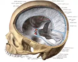
Falx cerebelli
The falx cerebelli is a small sickle-shaped fold of dura mater projecting forwards into the posterior cerebellar notch as well as projecting into the vallecula of the cerebellum between the two cerebellar hemispheres.[1]
| Falx cerebelli | |
|---|---|
 Falx cerebelli seen in back portion of skull. | |
 Occipital bone. Inner surface. (Portions "for faulx cerebelli" identified at center left.) | |
| Details | |
| Part of | Meninges |
| Identifiers | |
| Latin | Falx cerebelli |
| NeuroNames | 1238 |
| TA98 | A14.1.01.106 |
| TA2 | 5377 |
| FMA | 83974 |
| Anatomical terms of neuroanatomy | |
The name comes from two Latin words: falx, meaning "curved blade or scythe", and cerebellum, meaning "little brain".[2]
Anatomy
The falx cerebelli is a small midline fold of dura mater projecting anterior-ward from the skull and into the space between the cerebellar hemispheres.[3] It generally measures between 2.8 and 4.5 cm in length, and approximately 1–2 mm in thickness.[4]
Attachments
Superiorly, it (with its upwardly directed base) attaches at the midline to the posterior portion of the inferior surface of the tentorium cerebelli.[3]
Posteriorly, it attaches to the internal occipital crest; the inferior-most extremity of its posterior attachment frequently divides into two small folds that terminate at either side of the foramen magnum.[3]
Anatomical relations
The occipital sinus is contained within the posterior extremity of the falx cerebelli where it attaches to the internal occipital crest.[3]
Anatomical variation
In its lower portion the falx cerebelli diminishes very rapidly in height and as it descends, it can divide into two smaller folds or diverging limbs,[5] which are lost on the sides of the foramen magnum. Other variations such as duplication,[6] triplication,[7] absence,[8] and fenestration are much less common. As dural venous sinuses are concurrent with the development of dural folds, duplication of the falx cerebelli is usually associated with duplicated occipital sinus.[9] Knowledge of these variations is important in preventing iatrogenic injuries in this region.
See also
- Falx (disambiguation) — other parts of the anatomy with names including "falx"
References
- Atlas and textbook of human anatomy. Atlas der deskriptiven Anatomie des Menschen.English. Saunders. 1909.
- Sihler, Andrew L. (1995). New Comparative Grammar of Greek and Latin. Oxford University Press. ISBN 978-0-19-508345-3. Retrieved 12 March 2013.
- Standring, Susan (2021). Gray's Anatomy: The Anatomical Basis of Clinical Practice (42nd ed.). [New York]: Elsavier. ISBN 978-0-7020-7707-4. OCLC 1201341621.
- Shoja MM, Tubbs RS, Khaki AA, Shokouhi G. A rare variation of the posterior cranial fossa: duplicated falx cerebelli, occipital venous sinus, and internal occipital crest. Folia Morphol (Warsz) 2006;65(2):171–173.
- Atlas and textbook of human anatomy. Atlas der deskriptiven Anatomie des Menschen.English. Saunders. 1909.
- Shoja MM, Tubbs RS, Khaki AA, Shokouhi G. A rare variation of the posterior cranial fossa: duplicated falx cerebelli, occipital venous sinus, and internal occipital crest. Folia Morphol (Warsz) 2006;65(2):171–173.
- Shoja MM, Tubbs RS, Loukas M, Shokouhi G, Oakes WJ. A complex dural-venous variation in the posterior cranial fossa: a triplicate falx cerebelli and an aberrant venous sinus. Folia Morphol (Warsz) 2007;66(2):148–51.
- Tubbs Rs, Dockery SE, Salter G, Elton S, Blount JP, Grabb PA, Oakes WJ. Absence of the falx cerebelli in a Chiari II malformation. Clin Anat. 2002;15(3):193–195.
- Shoja MM, Tubbs RS, Shokouhi GH, Ashrafian A, Oakes WJ. Abstract presented at the 23rd Annual Meeting of the American Association of Clinical Anatomists. Milwaukee, Wisconsin: 2006. A triple dural-venous variation in the posterior cranial fossa: A duplicated plus accessory falx cerebelli and an aberrant venous sinus.
External links
- Anatomy photo:28:st-1601 at the SUNY Downstate Medical Center