Aortic bifurcation
The aortic bifurcation is the point at which the abdominal aorta bifurcates (forks) into the left and right common iliac arteries. The aortic bifurcation is usually seen at the level of L4,[1] just above the junction of the left and right common iliac veins.
| Aortic bifurcation | |
|---|---|
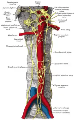
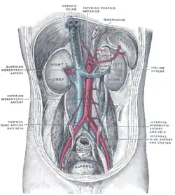
 The abdominal aorta and its bifurcation into the two common iliac arteries (red). | |
| Details | |
| Source | Abdominal aorta |
| Branches | Common iliac arteries |
| Vein | Inferior vena cava |
| Identifiers | |
| Latin | Bifurcatio aortae |
| TA98 | A12.2.13.001 |
| TA2 | 4297 |
| FMA | 3795 |
| Anatomical terminology | |
The right common iliac artery passes in front of the left common iliac vein. In some individual, mainly women with lumbar lordosis this vein can be compressed between the vertebra and the artery. This is the so-called Cockett syndrome or May–Thurner syndrome[2] can cause a slower venous flow and the possibility of deep venous thrombosis in the left leg mainly in pregnancy.
In surface anatomy, the bifurcation approximately corresponds to the umbilicus.[3]
Additional images
 The abdominal aorta and its branches.
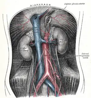
 Posterior abdominal wall, after removal of the peritoneum, showing kidneys, supra-renal capsules, and great vessels.
References
- Lerona PT, Tewfik HH (June 1975). "Bifurcation level of the aorta: landmark for pelvic irradiation". Radiology. 115 (3): 735. doi:10.1148/15.3.735. PMID 1129492.
- Becquemin JP, Juillet Y, Mexme M, Fiessinger JN, Cormier JM, Housset E (1981). "Clinical presentations of Cockett's syndrome". Nouv Presse Med. 70 (12): 959–62. PMID 7208320.
- Attwell, Lukas; Rosen, Sarah; Upadhyay, Bhavin; Gogalniceanu, Peter (5 June 2015). "The umbilicus: a reliable surface landmark for the aortic bifurcation?". Surgical and Radiologic Anatomy. 37 (10): 1239–1242. doi:10.1007/s00276-015-1500-1. PMID 26044782.
This article is issued from Wikipedia. The text is licensed under Creative Commons - Attribution - Sharealike. Additional terms may apply for the media files.