Claude's syndrome
| Claude's syndrome | |
|---|---|
 | |
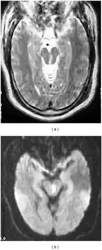
| a,b) Severely motion degraded axial T2-weighted and DWI images of midbrain demonstrate a subacute infarct within the left midbrain tegmentum | |
Claude's syndrome is a form of brainstem stroke syndrome characterized by the presence of an ipsilateral oculomotor nerve palsy, contralateral hemiparesis, contralateral ataxia, and contralateral hemiplegia of the lower face, tongue, and shoulder. Claude's syndrome affects oculomotor nerve, red nucleus and brachium conjunctivum.[1] Claude's syndrome is caused by midbrain infarction as a result of occlusion of a branch of the posterior cerebral artery.[2] This lesion is usually a unilateral infarction of the red nucleus and cerebellar peduncle, affecting several structures in the midbrain including:
| Structure damaged | Effect |
|---|---|
| dentatorubral tract fibers | contralateral ataxia |
| corticospinal tract fibers | contralateral hemiparesis |
| corticobulbar tract fibers | contralateral hemiplegia of lower facial muscles, tongue, and shoulder |
| oculomotor nerve fibers | ipsilateral oculomotor nerve palsy with a drooping eyelid and fixed wide pupil pointed down and out; probable diplopia |

It is very similar to Benedikt's syndrome.
It has been reported that posterior cerebral artery stenosis can also precipitate Claude's syndrome.[3]
It carries the name of Henri Charles Jules Claude, a French psychiatrist and neurologist, who described the condition in 1912.[4]
See also
- Wallenberg's syndrome
- Moritz Benedikt
References
- ↑ Harrison's
- ↑ "Claude's syndrome". GPnotebook.
- ↑ Dhanjal T, Walters M, MacMillan N (2003). "Claude's syndrome in association with posterior cerebral artery stenosis". Scottish Medical Journal. 48 (3): 91–92. doi:10.1177/003693300304800309. PMID 12968516. S2CID 7990190. Archived from the original on 2007-06-12.
- ↑ Claude H, Loyez M (1912). "Ramollissement du noyau rouge". Rev Neurol (Paris). 24: 49–51.
External links
| Classification |
|---|