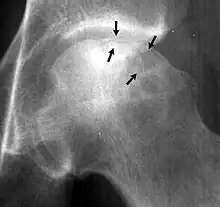
Crescent sign

In radiology, the crescent sign is a finding on conventional radiographs that is associated with avascular necrosis.[1][2][3] It usually occurs later in the disease, in stage III of the four-stage Ficat classification system.[1] It appears as a curved subchondral radiolucent line that is often found on the proximal femoral or humeral head.[1] Usually, this sign indicates a high likelihood of collapse of the affected bone.[1] The crescent sign may be best seen in an abducted (frog-legged) position.[1][4]
The crescent sign is caused by the necrotic and repair processes that occur during avascular necrosis.[1][2] Osteosclerosis occurs at a margin where new bone is placed over dead trabeculae.[1] When the trabeculae experience stress leading to microfractures and collapse, the crescent sign appears.[1]
The crescent sign may be seen with other bone diseases, such as shear fractures.[1]
References
- 1 2 3 4 5 6 7 8 9 Pappas, J. N. (2000). "The musculoskeletal crescent sign". Radiology. 217 (1): 213–214. doi:10.1148/radiology.217.1.r00oc22213. PMID 11012446.
- 1 2 Kenzora, J. E.; Glimcher, M. J. (1985). "Pathogenesis of idiopathic osteonecrosis: The ubiquitous crescent sign". The Orthopedic clinics of North America. 16 (4): 681–696. PMID 4058896.
- ↑ Norman, A.; Bullough, P. (1963). "The Radiolucent Crescent Line--An Early Diagnostic Sign of Avascular Necrosis of the Femoral Head". Bulletin of the Hospital for Joint Diseases. 24: 99–104. PMID 14048829.
- ↑ "Rheumatology Image Bank: Avascular Necrosis, Crescent Sign: Ficat Stage III, Hip". Retrieved 26 June 2012.