Intubation granuloma
| Intubation granuloma | |
|---|---|
 | |
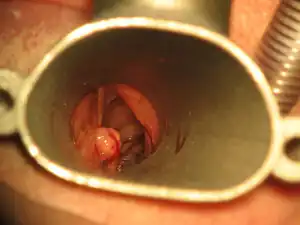
| Laryngoscopic view of the vocal process. An intubation granuloma is visible as a pale nodule on the left posterior laryngeal wall. |
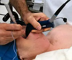
Intubation granuloma is a benign growth of granulation tissue in the larynx or trachea, which arises from tissue trauma due to endotracheal intubation.[1] This medical condition is described as a common late complication of tracheal intubation, specifically caused by irritation to the mucosal tissue of the airway during insertion or removal of the patient’s intubation tube.[1][2]
Endotracheal intubation is a common medical procedure, performed to assist patient ventilation and protect the airway.[2][3] However, prolonged endotracheal intubation, the use of inappropriate intubation equipment, or improper airway manipulation by the medical team may directly lead to mechanical trauma, resulting in laryngeal granuloma formation in the subglottis of the larynx.[4] Diagnosis of intubation granulomas are achieved through identifying proliferating tissues in the vocal folds via laryngoscopy.[2]
.jpg.webp)
Primary treatment for intubation granulomas tends to involve surgical excision of the granuloma. However, single treatment methods alone often result in high incidences of recurrence, hence combined therapy is suggested.[5] Secondary methods involve low dose radiotherapy and corticosteroid drug treatments.[6] For extreme cases of refractory granulomas, in which the aforementioned treatment methods all prove ineffective, botulinum toxin injections and oral zinc sulfate treatments are administered.[7][8]
Other significant risk factors are associated with intubation granuloma formation as well, such as a patient’s age, sex, intubation history and pre-existing medical conditions, which indirectly predispose certain patients to intubation-related injuries.[1][9]
Signs and Symptoms
Persistent sore throat, hoarseness, and vocal fatigue following intubation procedures are common symptoms of intubation granuloma, and patients may report mild discomfort associated with the sensation of a rough foreign body lodged in the back of the throat.[1][2][9] These symptoms often provoke observable clinical signs such as frequent coughing, throat-clearing, and hoarseness accompanied by dysphonia, reduced voice quality and restricted vocal range.[2][10] Severe intubation granulomas cause pharyngitis and pain upon pressed phonation, coughing or throat clearing.[11] In some cases, the patient may even experience dyspnea, or shortness of breath due to airway obstruction by the granuloma.[12][13]
However, since granulomas and other vocal cord polyps may take weeks or months to develop, intubation granulomas may sometimes be clinically evident only when the aforementioned symptoms persist for, or reappear after a longer period of time post-extubation.[14] Initial symptoms may also be overlooked as they coincide with typical side-effects of intubation.[15] Case reports of patients diagnosed and treated for intubation granulomas concur with this observation, as the diagnosis is often made weeks or months after the patient is extubated.[1][16]
Causes
Tracheal and laryngeal trauma leading to an intubation granuloma are caused by traumas during the intubation processes, directly resulting from technical circumstances such as specifications of the breathing tube equipment, method of insertion, and intubation duration.[4][9]

Intubation duration
Statistically, patients intubated for more than 48 hours will experience some form of laryngeal injury attributed to intubation, and approximately half of the injuries will result in the development of granulation tissue in the vocal fold.[17] While there is no consensus on the maximal permissible duration of safe endotracheal intubation, the risk of trauma-related laryngeal granuloma formation increases significantly with prolonged durations of tracheal intubation.[4][18] However, there are also studies which have not found statistically significant correlations between prolonged intubation duration with the degree of laryngeal injury, and intubation granuloma cases have also been reported in patients who have been intubated for only a few hours.[1][17]
Intubation tube diameter
Appropriate intubation tube sizes are defined as those small enough to minimise risks of mucosal trauma while large enough to maintain adequate ventilation.[4] This is especially important in the field of pediatrics, where the development of a child’s trachea may vary according to age.[19] Age-based calculations of appropriately sized intubation tubes are conducted in accordance with the Khine formula, which are based on internal diameters.[20] Unfortunately, these formulas do not account for variances in outer diameter and cuff dimensions, which may result in varying tube sizes.[21] Alternatively, height-based calculations are also available.[22] According to PALS (2010) guidelines, the use of length-based resuscitation tapes has proven to be more accurate than age-based estimates of endotracheal intubation tubes.[4]
Cuff pressure
The addition of an endotracheal tube cuff decreases the likelihood of selecting oversized breathing tubes for the patient, while also preventing microaspiration and the leakage of respiratory gases during intubation.[23] However, hyperinflation of the cuff places excessive pressure on the tracheal wall, causing trauma or ischaemia to nearby tissue and hence increasing the risk of granuloma formation.[24] Cuff pressures can be monitored during endotracheal intubation via manometers to prevent nitrous oxide induced hyperinflation.[25][26] General guidelines suggest that cuff pressure should be maintained between 20 to 30 cm to minimise risks of intubation-related trauma.[2][27]
Diagnosis
Intubation granulomas are most commonly presented in the form of red or pale spherical lesions in the subglottis of the larynx and may be defined as protruding, inflamed fibrovascular tissue.[10][17] While it is possible for intubation granulomas to form in both the larynx or trachea, they are most characteristically located in the posterior third aspect of the larynx, stemming from the posterior vocal fold directly above the vocal process cartilage.[28] Diagnosis of granulomas are confirmed via videolaryngostroboscopy and the electromyography by identifying proliferating tissue originating in the vocal process.[2] Furthermore, granuloma severity can be determined using screening images of laryngoscopy and graded in accordance to Farwell’s grading system.[29]
Pathophysiology
When a patient lies supine, the ventilation tube tends to rest on the posterior part of the larynx, above three major potential sites of damage: the arytenoid cartilage, posterior glottis, and cricoid cartilage.[30] Excessive pressure or friction from contact between the tube and the mucosal cell layer of the larynx, which may occur at rest or by unexpected myoclonic movement under sedation (such as coughing or swallowing), can lead to mucosal injury.[30] Under high capillary perfusion pressure, the mucosal cells of the larynx experience pressure ischemia, leading to tissue irritation, acute inflammation, congestion and edema.[30] Ischemic necrosis may occur, leading to erosion and ulcer formation in mucous membranes before progressing to the perichondrium and cartilage.[30] In other cases where granulomas are found in areas not on the posterior larynx, such tissue injury can also be accounted for by accidental lacerations from the tip of the endotracheal tube or its introducer.[30]
During prolonged intubation, constant stress on the laryngeal tissue prevents full wound recovery until the endotracheal tube is removed.[30] Although the formation of granulation tissue is part of a typical wound healing process, incomplete healing of the mucosal layer and persistent perichondritis causes the formation of chronic, rounded, localized granulation tissue over the ulceration site.[30] As the granulation tissue matures, other cells such as macrophages, fibroblasts and keratinocytes migrate to the granulation tissue to aid the healing process, causing fibrosis of the growth and the production of a protective epithelial layer.[31] Ultimately, a pedunculated globular mass consisting of immune cells, fibroblasts, myofibroblasts, keratinocytes and endothelial cells is formed.[31]
In some cases, the granuloma has been reported to regress after extubation without any medical intervention.[2] However, if the granuloma is not removed and continues to proliferate, this may pose further health risks to the patient, such as airway obstruction or stenosis.[2] In future intubations, even more caution would be required to perform the procedure while avoiding disruption of the granuloma.[13]
Treatment
The main treatment of intubation-related laryngeal granulomas is microlaryngeal surgical excision, but low dose radiotherapy and other drugs such as corticosteroids, botulinum toxin and zinc sulfate are also used in support to treat related symptoms or manage granuloma recurrence.[5][6]
Surgical excision
The main treatment of intubation-related laryngeal granulomas is microlaryngeal surgical excision of the granuloma under anesthesia.[28][6] Excision surgeries can be performed by cold steel excision or laser ablations - Laser surgeries permit more accurate excisions and hence reduce risks of damaging surrounding tissues.[32] This method can be further accompanied by jet ventilation, which minimises intubation trauma and reduces risks of edema and barotrauma by providing ventilation over stenosis.[28] A thin cannula and catheter can be further used in place of traditional small-diameter endotracheal tubes during surgery, which enables precise visualisation of anatomical configurations within the surgical field.[28] Employing infraglottic transtracheal routes for microlaryngeal surgery is more effective than supraglottic methods as it provides ventilation under vocal cords, which causes minimal vocal cord movement.[33]
However, excision surgeries alone usually result in high incidences of granuloma recurrence.[5] Consequently, surgical approaches are usually accompanied by low dose radiotherapy, corticosteroids and botulinum toxin treatment.[7][8][34][35]
Low dose radiotherapy
Low dose radiotherapy ranging between 800 to 3000 cGy (centigray) has been documented to have a high successful prevention and resolution of laryngeal granulomas.[35] The optimal period for radiotherapy treatment is immediately after surgical excision, preferably prior to injury-stimulated tissue proliferation.[36]
Corticosteroids
Corticosteroid drug treatments can be administered orally and through inhalation. Inhaled steroids have the greatest efficacy in resolving reducing local inflammation of the granuloma.[11][34][37] The most commonly prescribed inhaled steroid, budesonide, can resolve intubation granulomas within 12 months of treatment.[34]
However, due to the side effects of steroidal interventions, antibiotics have to be prescribed alongside to reduce pain and inflammation in the region of the target granuloma.[37]
Botulinum toxin and Zinc sulfate
Botulinum toxin (BOTOX) and Zinc sulfate treatments are mainly applied to cases of refractory granulomas, which are immune to previously mentioned treatment methods.[7][8]
Intralaryngeal BOTOX injections bind specifically and non-competitively to presynaptic cholinergic neuron membranes at neuromuscular junctions which induce zinc-dependent cleavage of proteins involved in neuroexocytosis.[38] The breakdown of neuroexocytosis proteins block acetylcholine secretions which inhibit hypertonicity, strengthen antagonist muscles and restore the balance of forces.[38] Since laryngeal granuloma formations are exacerbated by repeated forceful contraction of the glottis, the combined effects of the toxin induce thyroarytenoid paresis and decreases the force of vocal fold adduction which inhibit forced contact between vocal processes, hence facilitating granuloma resolution.[39][40]
Oral zinc sulfate treatments are advantageous due to their ability to preserve the anatomical and functional integrity of the vocal cords.[8] Similarly, this form of therapy can achieve quick relief of granuloma-related symptoms whilst avoiding invasive surgery and toxic drug effects.[8]
Epidemiology
Intubation granuloma onset has been found to be more prevalent in certain demographics due to their associated anatomical characteristics.[9] The physiological differences due to age, gender, or inherited features may place such patients at an increased risk of intubation injury, and subsequently the occurrence of intubation granulomas.[4][9]
Age
Pediatric and geriatric patients are at higher risk of laryngeal injury.[4] Compared to adults, newborns and young children possess a higher, more anterior larynx, a larger and stiffer epiglottis as well as a more fragile laryngotracheal mucosa, making them more vulnerable to traumatic damage by prolonged tracheal intubation.[4][9] In addition, the fragility of the mucous larynx increases with age, leaving the patient more prone to intubation-induced tracheal and laryngeal injuries.[2][41]
Gender
Females were found to be at greater risk of intubation granulomas as they tend to have a narrower glottis, lower glottic proportion and a thinner arytenoid mucochondrium.[1][9] 75% to 90% of intubation granulomas found in the vocal cords are reported in female patients.[1][34] Furthermore, females displayed greater postintubation pharyngitis, which have led to increased incidence of intubation granulomas.[42]
Anatomical characteristics
Congenital and/or acquired abnormalities of the larynx - laryngeal webs, bands, cysts and tumours - are predisposing risk factors of intubation granuloma.[9] In addition, facial and cervical anomalies, short necks, receding chins and obesity can heighten the difficulty in successful laryngoscopy, predisposing the patient to traumatic intubation as their airway becomes more challenging to navigate during the intubation process.[1][9]
References
- 1 2 3 4 5 6 7 8 9 Park, Si-Yeon; Choi, Hong Seok; Yoon, Ji-Young; Kim, Eun-Jung; Yoon, Ji-Uk; Kim, Hee Young; Ahn, Ji-Hye (December 2018). "Fatal vocal cord granuloma after orthognathic surgery". Journal of Dental Anesthesia and Pain Medicine. 18 (6): 375–378. doi:10.17245/jdapm.2018.18.6.375. ISSN 2383-9309. PMC 6323036. PMID 30637348.
- 1 2 3 4 5 6 7 8 9 10 Mota, Luiz; de Cavalho, Glauber; Brito, Valeska (April 2012). "Laryngeal complications by orotracheal intubation: Literature review". International Archives of Otorhinolaryngology. 16 (2): 236–245. doi:10.7162/S1809-97772012000200014. ISSN 1809-9777. PMC 4399631. PMID 25991942.
- ↑ Stone, Shepard B. (2007-01-01), Dehn, Richard W.; Asprey, David P. (eds.), "Chapter 12 - Endotracheal Intubation", Essential Clinical Procedures (Second Edition), W.B. Saunders, pp. 145–164, doi:10.1016/b978-1-4160-3001-0.50016-2, ISBN 978-1-4160-3001-0, retrieved 2020-04-27
- 1 2 3 4 5 6 7 8 Jang, Minyoung; Basa, Krystyne; Levi, Jessica (April 2018). "Risk factors for laryngeal trauma and granuloma formation in pediatric intubations". International Journal of Pediatric Otorhinolaryngology. 107: 45–52. doi:10.1016/j.ijporl.2018.01.008. ISSN 0165-5876. PMID 29501310.
- 1 2 3 Wu, Jingyi; Jiang, Tongchao; Wu, Yu; Ding, Lijuan; Dong, Lihua (2019-09-27). "Laryngeal granuloma occurring after surgery for laryngeal cancer treated by surgical removal and immediate post-operative radiotherapy". Medicine. 98 (39): e17345. doi:10.1097/MD.0000000000017345. ISSN 0025-7974. PMC 6775417. PMID 31574876.
- 1 2 3 Rimoli, Caroline Fernandes; Martins, Regina Helena Garcia; Catâneo, Daniele Cristina; Imamura, Rui; Catâneo, Antonio José Maria; Rimoli, Caroline Fernandes; Martins, Regina Helena Garcia; Catâneo, Daniele Cristina; Imamura, Rui; Catâneo, Antonio José Maria (December 2018). "Treatment of post-intubation laryngeal granulomas: systematic review and proportional meta-analysis". Brazilian Journal of Otorhinolaryngology. 84 (6): 781–789. doi:10.1016/j.bjorl.2018.03.003. ISSN 1808-8694. PMID 29699879.
- 1 2 3 Fink, Daniel S.; Achkar, Jihad; Franco, Ramon A.; Song, Phillip C. (2013-09-20). "Interarytenoid botulinum toxin injection for recalcitrant vocal process granuloma". The Laryngoscope. 123 (12): 3084–3087. doi:10.1002/lary.23915. ISSN 0023-852X. PMID 24115127. S2CID 9919682.
- 1 2 3 4 5 Djukić, Vojko; Krejović-Trivić, Sanja; Vukašinović, Milan; Trivić, Aleksandar; Pavlović, Bojan; Milovanović, Aleksandar; Milovanović, Jovica (April 2015). "Laryngeal Granuloma – Benefit in Treatment with Zinc Supplementation?". Journal of Medical Biochemistry. 34 (2): 228–232. doi:10.2478/jomb-2014-0028. ISSN 1452-8258. PMC 4922326. PMID 28356836.
- 1 2 3 4 5 6 7 8 9 Blanc, V. F.; Tremblay, N. A. (March 1974). "The complications of tracheal intubation: a new classification with a review of the literature". Anesthesia and Analgesia. 53 (2): 202–213. doi:10.1213/00000539-197403000-00005. ISSN 0003-2999. PMID 4593090. S2CID 42218487.
- 1 2 "Granuloma | Sean Parker Institute for the Voice". voice.weill.cornell.edu. Retrieved 2020-04-08.
- 1 2 Martins, Regina Helena Garcia; Braz, José Reinaldo Cerqueira; Dias, Norimar Hernandes; Castilho, Emanuel Celice; Braz, Leandro Gobbo; Navarro, Lais Helena Camacho (April 2006). "Hoarseness after tracheal intubation". Revista Brasileira de Anestesiologia. 56 (2): 189–199. doi:10.1590/S0034-70942006000200011. ISSN 0034-7094. PMID 19468566.
- ↑ "Contact Granulomas: Background, Problem, Etiology". 2020-02-19.
{{cite journal}}: Cite journal requires|journal=(help) - 1 2 Nakahira, Junko; Sawai, Toshiyuki; Matsunami, Sayuri; Minami, Toshiaki (December 2014). "Worst-case scenario intubation of laryngeal granuloma: a case report". BMC Research Notes. 7 (1): 74. doi:10.1186/1756-0500-7-74. ISSN 1756-0500. PMC 3937148. PMID 24490715.
- ↑ Kacmarek, Robert M.; Stoller, James K.; Heuer, Al (2016-02-05). Egan's Fundamentals of Respiratory Care - E-Book. Elsevier Health Sciences. ISBN 978-0-323-39385-0.
- ↑ Cho, Choon-Kyu; Kim, Jae-Jung; Sung, Tae-Yun; Jung, Sung-Mee; Kang, Po-Soon (December 2013). "Endotracheal intubation-related vocal cord ulcer following general anesthesia". Korean Journal of Anesthesiology. 65 (6 Suppl): S147–S148. doi:10.4097/kjae.2013.65.6S.S147. ISSN 2005-6419. PMC 3903841. PMID 24478853.
- ↑ Song, Jae Gyok; Cho, Won Ho; Ji, Sung Mi; Park, Jeong Heon; Kim, Seok Kon (2019-10-31). "Laryngeal granulomas in patients after two-jaw surgery - Four cases report -". Anesthesia and Pain Medicine. 14 (4): 489–493. doi:10.17085/apm.2019.14.4.489. ISSN 1975-5171. PMC 7713798. PMID 33329782.
- 1 2 3 Colton House, Joyce; Noordzij, J. Pieter; Murgia, Bobby; Langmore, Susan (2010-12-16). "Laryngeal injury from prolonged intubation: A prospective analysis of contributing factors". The Laryngoscope. 121 (3): 596–600. doi:10.1002/lary.21403. ISSN 0023-852X. PMC 3084628. PMID 21344442.
- ↑ KASTANOS, NIKOS; MIRÓ, RAMON ESTOPÁ; PEREZ, ALBERTO MARÍN; MIR, ANTONIO XAUBET; AGUSTÍ-VIDAL, ALBERTO (May 1983). "Laryngotracheal injury due to endotracheal intubation". Critical Care Medicine. 11 (5): 362–367. doi:10.1097/00003246-198305000-00009. ISSN 0090-3493. PMID 6839788. S2CID 31803648.
- ↑ Kim, Hee Young; Cheon, Ji Hyun; Baek, Seung Hoon; Kim, Kyung Hoon; Kim, Tae Kyun (February 2017). "Prediction of endotracheal tube size for pediatric patients from the epiphysis diameter of radius". Korean Journal of Anesthesiology. 70 (1): 52–57. doi:10.4097/kjae.2017.70.1.52. ISSN 2005-6419. PMC 5296388. PMID 28184267.
- ↑ Weiss, Markus; Gerber, Andreas (November 2008). "Evaluation of cuffed tracheal tube size predicted using the Khine formula in children". Pediatric Anesthesia. 18 (11): 1105. doi:10.1111/j.1460-9592.2008.02676.x. ISSN 1155-5645. PMID 18950336. S2CID 205519435.
- ↑ Weiss, M. Dullenkopf, A. Gysin, C. Dillier, C. M. Gerber, A. C. Shortcomings of cuffed paediatric tracheal tubes†. OCLC 999833677.
{{cite book}}: CS1 maint: multiple names: authors list (link) - ↑ Sutagatti, Jagadish G; Raja, Ranjana; Kurdi, Madhuri S (May 2017). "Ultrasonographic Estimation of Endotracheal Tube Size in Paediatric Patients and its Comparison with Physical Indices Based Formulae: A Prospective Study". Journal of Clinical and Diagnostic Research. 11 (5): UC05–UC08. doi:10.7860/JCDR/2017/25905.9838. ISSN 2249-782X. PMC 5483782. PMID 28658880.
- ↑ Hamilton, V. Anne; Grap, Mary Jo (March 2012). "The role of the endotracheal tube cuff in microaspiration". Heart & Lung. 41 (2): 167–172. doi:10.1016/j.hrtlng.2011.09.001. ISSN 0147-9563. PMC 3828744. PMID 22209048.
- ↑ Henderson, John (2010), "Airway Management in the Adult", Miller's Anesthesia, Elsevier, pp. 1573–1610, doi:10.1016/b978-0-443-06959-8.00050-9, ISBN 978-0-443-06959-8
- ↑ Trivedi, Lopa; Jha, Pramila; Bajiya, NarasiRam; Tripathi, DC (2010). "We should care more about intracuff pressure: The actual situation in government sector teaching hospital". Indian Journal of Anaesthesia. 54 (4): 314–7. doi:10.4103/0019-5049.68374. ISSN 0019-5049. PMC 2943700. PMID 20882173.
- ↑ Kumar, Chandra M.; Seet, Edwin; Van Zundert, Tom C. R. V. (2020-03-20). "Measuring endotracheal tube intracuff pressure: no room for complacency". Journal of Clinical Monitoring and Computing. 35 (1): 3–10. doi:10.1007/s10877-020-00501-2. ISSN 1387-1307. PMC 7223496. PMID 32198671.
- ↑ MACKENZIE, C.F.; KLOSE, S.; BROWNE, D.R.G. (February 1976). "A STUDY OF INFLATABLE CUFFS ON ENDOTRACHEAL TUBES: Pressures exerted on the trachea". British Journal of Anaesthesia. 48 (2): 105–110. doi:10.1093/bja/48.2.105. ISSN 0007-0912. PMID 766796.
- 1 2 3 4 Altun, Demet; Yılmaz, Eren; Başaran, Bora; Çamcı, Emre (August 2014). "Surgical Excision of Postintubation Granuloma Under Jet Ventilation". Turkish Journal of Anaesthesiology and Reanimation. 42 (4): 220–222. doi:10.5152/TJAR.2014.16362. ISSN 2149-0937. PMC 4894151. PMID 27366423.
- ↑ Farwell, D G; Belafsky, P C; Rees, C J (2008-03-03). "An endoscopic grading system for vocal process granuloma". The Journal of Laryngology & Otology. 122 (10): 1092–1095. doi:10.1017/s0022215108001722. ISSN 0022-2151. PMID 18312706. S2CID 11134790.
- 1 2 3 4 5 6 7 Benjamin, Bruce (2018-07-17). "Prolonged Intubation Injuries of the Larynx: Endoscopic Diagnosis, Classification, And Treatment". Annals of Otology, Rhinology & Laryngology. 127 (8): 492–507. doi:10.1177/0003489418790348. ISSN 0003-4894. PMID 30012012. S2CID 51638585.
- 1 2 Alhajj, Mandy; Bansal, Pankaj; Goyal, Amandeep (2020), "Physiology, Granulation Tissue", StatPearls, StatPearls Publishing, PMID 32119289, retrieved 2020-04-08
- ↑ Karkos, Petros D.; George, Michael; Van Der Veen, Jan; Atkinson, Helen; Dwivedi, Raghav C.; Kim, Dae; Repanos, Costa (2014-03-17). "Vocal Process Granulomas". Annals of Otology, Rhinology & Laryngology. 123 (5): 314–320. doi:10.1177/0003489414525921. ISSN 0003-4894. PMID 24642585. S2CID 45781990.
- ↑ Hunsaker, D. H. (August 1994). "Anesthesia for microlaryngeal surgery: the case for subglottic jet ventilation". The Laryngoscope. 104 (8 Pt 2 Suppl 65): 1–30. doi:10.1002/lary.1994.104.s65.1. ISSN 0023-852X. PMID 8052087. S2CID 22778328.
- 1 2 3 4 D., Wael; Fathy, Essam; Attya, Sameer; ELSHABBOURY, MOHAMMED (2017-11-01). "Steroid Inhalation Versus Surgery in Treatment of Post-Intubation Granuloma". Zagazig University Medical Journal. 21 (6): 1–5. doi:10.21608/zumj.2017.4578. ISSN 2357-0717.
- 1 2 Mitchell, G.; Pearson, C. R.; Henk, J. M.; Rhys-Evans, P. (May 1998). "Excision and low-dose radiotherapy for refractory laryngeal granuloma". The Journal of Laryngology & Otology. 112 (5): 491–493. doi:10.1017/s0022215100140873. ISSN 0022-2151. PMID 9747485.
- ↑ Harari, P. M.; Blatchford, S. J.; Coulthard, S. W.; Cassady, J. R. (May 1991). "Intubation granuloma of the larynx: successful eradication with low-dose radiotherapy". Head & Neck. 13 (3): 230–233. doi:10.1002/hed.2880130312. ISSN 1043-3074. PMID 2037475. S2CID 11589561.
- 1 2 Hoffman, H. T.; Overholt, E.; Karnell, M.; McCulloch, T. M. (December 2001). "Vocal process granuloma". Head & Neck. 23 (12): 1061–1074. doi:10.1002/hed.10014. ISSN 1043-3074. PMID 11774392. S2CID 33265680.
- 1 2 Orloff, Lisa A.; Goldman, Stephen N. (September 1999). "Vocal Fold Granuloma: Successful Treatment with Botulinum Toxin". Otolaryngology–Head and Neck Surgery. 121 (4): 410–413. doi:10.1016/s0194-5998(99)70230-5. ISSN 0194-5998. PMID 10504597. S2CID 7837798.
- ↑ Nasri, Sina; Sercarz, Joel A.; Mcalpin, Tina; Berke, Gerald S. (June 1995). "Treatment of vocal fold granuloma using botulinum toxin type A". The Laryngoscope. 105 (6): 585–588. doi:10.1288/00005537-199506000-00005. ISSN 0023-852X. PMID 7769940. S2CID 19392973.
- ↑ Lemos, Elza Maria; Sennes, Luiz Ubirajara; Imamura, Rui; Tsuji, Domingos H. (August 2005). "Vocal process granuloma: clinical characterization, treatment and evolution". Revista Brasileira de Otorrinolaringologia. 71 (4): 494–498. doi:10.1590/S0034-72992005000400016. ISSN 0034-7299. PMID 16446966.
- ↑ Bhardwaj, Neerja (2013). "Pediatric cuffed endotracheal tubes". Journal of Anaesthesiology Clinical Pharmacology. 29 (1): 13–18. doi:10.4103/0970-9185.105786. ISSN 0970-9185. PMC 3590525. PMID 23492803.
- ↑ El‐Boghdadly, K.; Bailey, C. R.; Wiles, M. D. (2016). "Postoperative sore throat: a systematic review". Anaesthesia. 71 (6): 706–717. doi:10.1111/anae.13438. ISSN 1365-2044. PMID 27158989. S2CID 25837446.