Blue nevus
| Blue nevus | |
|---|---|
| Other names: Dendritic blue nevus, common blue nevus, dermal denf=dritic melanocytotic nevus, nevus of Jadassohn, Tièche nevus, Jadassohn-Tièche nevus[1] | |
.jpg.webp) | |
| Blue nevus | |
| Specialty | Dermatology |
| Symptoms | Single well-defined blue-black bump[1] |
| Complications | Rarely malignant transformation[2] |
| Types | Dendritic, cellular[1] |
| Causes | Unclear[2] |
| Diagnostic method | Visualisation, dermoscopy[3] |
| Differential diagnosis | Dermatofibroma, melanoma[2][4] |
| Treatment | Monitoring, excision[2] |
| Prognosis | Good[2] |
| Frequency | Female>male[5] |
A blue nevus is a type of coloured mole, typically a single well-defined blue-black bump.[1]
The blue colour is caused by the pigment being deep in the skin.[3]
Diagnosis is by visualisation and dermoscopy.[3] A biopsy is sometimes performed, or the whole lesion surgically removed.[2] The outcome is generally good but there is a small chance of cancerous transformation.[2] Differential diagnosis includes dermatofibroma and melanoma.[2]
Blue nevi are more common in females than males.[5] It was first studied in 1906 by Tièche, a student of Josef Jadassohn.[6]
Classification
Blue nevi may be divided into the following types:[7]: 701
- A patch blue nevus (also known as an "acquired dermal melanocytosis", and "dermal melanocyte hamartoma") is a cutaneous condition characterized by a diffusely gray-blue area that may have superimposed darker macules.[8]
- A blue nevus of Jadassohn–Tièche (also known as a "common blue nevus", and "nevus ceruleus") is a cutaneous condition characterized by a steel-blue papule or nodule.[7]: 701
- A cellular blue nevus is a cutaneous condition characterized by large, firm, blue or blue-black nodules.[7]: 701
- An epithelioid blue nevus is a cutaneous condition most commonly seen in patients with the Carney complex.[7]: 701
- A deep penetrating nevus is a type of benign melanocytic skin tumor characterized, as its name suggests, by penetration into the deep dermis and/or subcutis. Smudged chromatic is a typical finding. In some cases mitotic figures or atypical melanocytic cytology are seen, potentially mimicking a malignant melanoma. Evaluation by an expert skin pathologist is advisable in some cases to help differentiate from invasive melanoma.[7]: 701
- An amelanotic blue nevus (also known as a "hypomelanotic blue nevus") is a cutaneous condition characterized by mild atypia and pleomorphism.[7]: 701
- A malignant blue nevus is a cutaneous condition characterized by a sheet-like growth pattern, mitoses, necrosis, and cellular atypia.[8][7]: 701
Signs and symptoms
It is typically a single well-defined blue-black bump.[5]
.jpg.webp) Blue nevus
Blue nevus.jpg.webp) Blue naevus
Blue naevus.jpg.webp) Blue naevus
Blue naevus
Diagnosis
Diagnosis is by visualisation and dermoscopy.[3] A biopsy is sometimes performed.[2]
Dermoscopy
.jpg.webp) Blue naevus
Blue naevus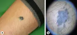 Blue nevus and dermoscopy
.jpg.webp)
Histopathology
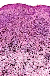 Micrograph of a blue nevus showing the characteristic pigmented melanocytes between bundles of collagen. H&E stain.
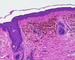 Blue nevus
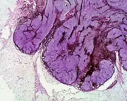 Cellular blue nevus
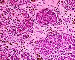 Epithelioid blue nevus
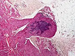 Malignant blue nevus
Epidemiology

Blue nevi are more common in females than males.[5]
History
It was first studied in 1906 by Tièche, a student of Josef Jadassohn.[6]
See also
References
- 1 2 3 4 DE, Elder; D, Massi; RA, Scolyer; R, Willemze (2018). "2. Melanocytic tumours: Blue nevus and cellular blue naevus". WHO Classification of Skin Tumours. Vol. 11 (4th ed.). Lyon (France): World Health Organization. pp. 126–129. ISBN 978-92-832-2440-2. Archived from the original on 2022-07-11. Retrieved 2022-08-20.
- 1 2 3 4 5 6 7 8 9 Austad, Steve S.; Athalye, Leela (2021). "Blue Nevus". StatPearls. StatPearls Publishing. Archived from the original on 2021-10-13. Retrieved 2021-10-12.
- 1 2 3 4 "Blue naevus". dermnetnz.org. Archived from the original on 13 August 2021. Retrieved 12 October 2021.
- ↑ Blue+Nevi at the US National Library of Medicine Medical Subject Headings (MeSH)
- 1 2 3 4 Johnstone, Ronald B. (2017). "32. Lentigines and melanomas". Weedon's Skin Pathology Essentials (2nd ed.). Elsevier. p. 545. ISBN 978-0-7020-6830-0. Archived from the original on 2021-05-25. Retrieved 2021-09-25.
- 1 2 Sreeremya, S. (17 April 2018). "Blue Nevus". International Journal of Molecular Biotechnology. 4 (1): 1–4. doi:10.37628/ijmb.v4i1.255. Archived from the original on 13 October 2021. Retrieved 12 October 2021.
- 1 2 3 4 5 6 7 James, William D.; Berger, Timothy G.; et al. (2006). Andrews' Diseases of the Skin: clinical Dermatology. Saunders Elsevier. ISBN 0-7216-2921-0.
- 1 2 Rapini, Ronald P.; Bolognia, Jean L.; Jorizzo, Joseph L. (2007). Dermatology: 2-Volume Set. St. Louis: Mosby. p. 1722. ISBN 978-1-4160-2999-1.
External links
| Classification |
|---|