Meibomian gland
| Meibomian gland | |
|---|---|
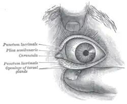
 Front of left eye with eyelids separated to show medial canthus and openings of meibomian (tarsal) glands | |
| Details | |
| System | Integumentary |
| Identifiers | |
| Latin | glandulae tarsales |
| MeSH | D008537 |
| TA98 | A15.2.07.042 |
| TA2 | 6833 |
| FMA | 71872 |
| Anatomical terminology | |
Meibomian glands (also called tarsal glands, palpebral glands, and tarsoconjunctival glands) are sebaceous glands along the rims of the eyelid inside the tarsal plate. They produce meibum, an oily substance that prevents evaporation of the eye's tear film. Meibum prevents tears from spilling onto the cheek, traps them between the oiled edge and the eyeball, and makes the closed lids airtight.[1] There are about 25 such glands on the upper eyelid, and 20 on the lower eyelid.
Dysfunctional meibomian glands is believed to be the most often cause of dry eyes. They are also the cause of posterior blepharitis.[2]
History

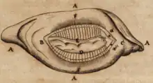
The glands were mentioned by Galen in 200 AD[3] and were described in more detail by Heinrich Meibom (1638–1700), a German physician, in his work De Vasis Palpebrarum Novis Epistola in 1666. This work included a drawing with the basic characteristics of the glands.[4][5]
Anatomy
Although the upper lid have greater number and volume of meibomian glands than the lower lid, there is no consensus whether it contributes more to the tearfilm stability. The glands do not have direct contact with eyelash follicles. The process of blinking releases meibum into the lid margin. [2]
Function
Meibum
Lipids
Lipids are the major components of meibum (also known as "meibomian gland secretions"). The term "meibum" was originally introduced by Nicolaides et al. in 1981.[6]
The biochemical composition of meibum is extremely complex and very different from that of sebum. Lipids are universally recognized as major components of human and animal meibum. An update was published in 2009 on the composition of human meibum and on the structures of various positively identified meibomian lipids[7]
Currently, the most sensitive and informative approach to lipidomic analysis of meibum is mass spectrometry, either with direct infusion[8][9] or in combination with liquid chromatography.[10]
The lipids are the main component of the lipid layer of the tear film, preventing rapid evaporation and it is believed they lower the surface tension help helps to stabilize the tear film.[3]
Proteins
In humans, more than 90 different proteins have been identified in meibomian gland secretions.[11]
Clinical significance
![Meibomian glands in the lower eyelid imaged under amber light to show vasculature support and the gland structure [epiCam].](../I/Meibomian-glands.png.webp)
Dysfunctional meibomian glands often cause dry eyes, one of the more common eye conditions. They may also contribute to blepharitis. Inflammation of the meibomian glands (also known as meibomitis, meibomian gland dysfunction, or posterior blepharitis) causes the glands to be obstructed by thick, cloudy-to-yellow, more opaque and viscous-like, oily and waxy secretions, a change from the glands' normal clear secretions.[12][13] Besides leading to dry eyes, the obstructions can be degraded by bacterial lipases, resulting in the formation of free fatty acids, which irritate the eyes and sometimes cause punctate keratopathy.
Meibomian gland dysfunction is more often seen in women, and is regarded as the main cause of dry eye disease.[14][15] Factors that contribute to meibomian gland dysfunction can include things such as a person's age and/or hormones,[16] or severe infestation of Demodex brevis mite.
Treatment can include warm compresses to thin the secretions and eyelid scrubs with baby shampoo or eyelid cleanser,[17][13] or emptying ("expression") of the gland by a professional. Lifitegrast and Restasis are topical medication commonly used to control the inflammation and improve the oil quality. In some cases topical steroids and topical (drops or ointment)/oral antibiotics (to reduce bacteria on the lid margin) are also prescribed to reduce inflammation.[13] Intense pulsed light (IPL) treatments have also been shown to reduce inflammation and improve gland function. Meibomian gland probing is also used on patients who experience deep clogging of the glands.
Meibomian gland dysfunction may be caused by some prescription medications, notably isotretinoin. A blocked meibomian gland can cause a chalazion (or "meibomian cyst") to form in the eyelid.
See also
References
- ↑ "eye, human." Encyclopædia Britannica. Encyclopædia Britannica Ultimate Reference Suite. Chicago: Encyclopædia Britannica, 2010.
- 1 2 Kelly K. Nichols; Gary N. Foulks; Anthony J. Bron; Ben J. Glasgow; Murat Dogru; Kazuo Tsubota; Michael A. Lemp; David A. Sullivan (2011). "The International Workshop on Meibomian Gland Dysfunction: Executive Summary". IOVS. 52 (4): 1922–1929. doi:10.1167/iovs.10-6997a. PMC 3072157. PMID 21450913.
{{cite journal}}: CS1 maint: uses authors parameter (link) - 1 2 Knop, Erich; Knop, Nadja; Millar, Thomas; Obata, Hiroto; Sullivan, David A. (2011). "The International Workshop on Meibomian Gland Dysfunction: Report of the Subcommittee on Anatomy, Physiology, and Pathophysiology of the Meibomian Gland". Investigative Ophthalmology & Visual Science. 52 (4): 1938–1978. doi:10.1167/iovs.10-6997c. PMC 3072159. PMID 21450915.
- ↑ "Meibomian Gland Dysfunction (MGD) - EyeWiki". eyewiki.aao.org.
- ↑ Meibomii, Henrici (1666). De vasis palpebrarum novis epistola … [From a recent letter on the eyelids' vesicles to the most renowned gentleman Dr. Joel Langelott, court physician of the most reverend and serene Duke of Holstein] (in Latin). Helmstadt, (Germany): Henning Muller.
- ↑ Nicolaides, N; Kaitaranta, JK; Rawdah, TN; Macy, JI; Boswell, FM; Smith, RE (April 1981). "Meibomian gland studies: comparison of steer and human lipids". Invest Ophthalmol Vis Sci. 20 (4): 522–536. PMID 7194326.
- ↑ Butovich IA (November 2009). "The Meibomian puzzle: combining pieces together". Prog Retin Eye Res. 28 (6): 483–498. doi:10.1016/j.preteyeres.2009.07.002. PMC 2783885. PMID 19660571.
- ↑ Chen, J; Green-Church, K; Nichols, K (2010). "Shotgun lipidomic analysis of human meibomian gland secretions with electrospray ionization tandem mass spectrometry". Invest Ophthalmol Vis Sci. 51 (12): 6220–6231. doi:10.1167/iovs.10-5687. PMC 3055753. PMID 20671273.
- ↑ Chen, J; Nichols, K (2018). "Comprehensive shotgun lipidomics of human meibomian gland secretions using MS/MSall with successive switching between acquisition polarity modes". J Lipid Res. 59 (11): 2223–2236. doi:10.1194/jlr.D088138. PMC 6210907. PMID 30279222.
- ↑ Butovich, IA (2009). Lipidomic analysis of human meibum using HPLC-MSn. Methods Mol Biol. Vol. 579. pp. 221–246. doi:10.1194/jlr.D088138. PMID 19763478.
- ↑ Tsai, PS; Evans, JE; Green, KM; Sullivan, RM; Schaumberg, DA; Richards, SM; Dana, MR; Sullivan, DA (March 2006). "Proteomic analysis of human meibomian gland secretions". Br J Ophthalmol. 90 (3): 372–7. doi:10.1136/bjo.2005.080846. PMC 1856970. PMID 16488965.
- ↑ Chhadva, Priyanka; Goldhardt, Raquel; Galor, Anat (24 November 2017). "Meibomian gland disease: the role of gland dysfunction in dry eye disease". Ophthalmology. 124 (11 Suppl): S20–S26. doi:10.1016/j.ophtha.2017.05.031. PMC 5685175. PMID 29055358.
- 1 2 3 Peter Bex, Reza Dana, Linda Mcloon, Jerry Niederkorn (2011). Ocular Periphery and Disorders
- ↑ "Managing and Making Sense of MGD". Review of Ophthalmology. 2012. Retrieved 26 February 2014.
- ↑ "Rethinking Meibomian Gland Dysfunction: How to Spot It, Stage It and Treat It". American Academy of Ophthalmology. 2014. Retrieved 26 February 2014.
- ↑ "The Role of Meibomian Gland Dysfunction and Lid Wiper Epitheliopathy in Dry Eye Disease". American Academy of Optometry. 2012. Archived from the original on 9 October 2013. Retrieved 26 February 2014.
- ↑ Aryasit, Orapan; Uthairat, Yuwarat; Singha, Penny; Horatanaruang, Orasa (8 May 2020). "Efficacy of baby shampoo and commercial eyelid cleanser in patients with meibomian gland dysfunction". Medicine. 99 (19): e20155. doi:10.1097/MD.0000000000020155. PMC 7220370. PMID 32384504.