Common mole (medicine)
| Common mole (medicine) | |
|---|---|
| Other names: Banal nevus, common acquired melanocytic nevus, nevocellular nevus, nevocytic nevus | |
 | |
| Compound nevus type | |
| Specialty | Dermatology[1] |
| Types | Junctional, compound, dermal[2] |
| Differential diagnosis | Melanoma[2] |
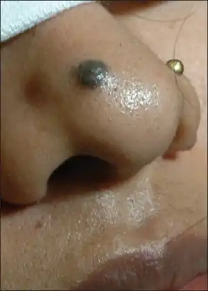
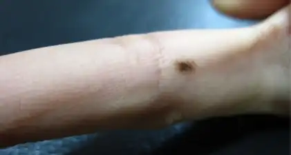
A common mole, also known as benign melanocytic nevus is a noncancerous pigmented spot or small bump in skin.[1] They are described as either junctional, compound, or dermal, and are typically less than 5mm in diameter, and mostly symmetrical and uniform in color.[2] Broadly; junctional nevus is a flat brown-black spot, dermal nevus is light or fleshy-colored and slightly elevated, and compound nevus is fleshy-colored to brown domes which sometimes appear as though it may fall off.[3]
It is generally acquired during childhood and reach a maximum number in young adults.[2] The skin cancer melanoma can appear similar.[2]
Types
The three most common categories of benign moles are those located at the border between the epidermis and dermis (junctional nevi), dermal nevi in the dermis only, and those found in both the dermis and epidermis (compound nevi).[1]
Junctional nevus
Junctional nevus appears as a brown to black well-defined flat marking in skin, typically appearing in childhood and adolescents; 3 to 18 years of age.[1] It can become a dermal or compound nevus later on.[1] In a nail it appears as a longitudinal line.[3]
Dermal nevus
Dermal nevus, also known as intradermal nevus, is a light or fleshy-colored small bump in skin, typically occurring in adults.[3] It may be a larger bump and have a protruding hair.[3] It is the commonest type of melanocytic nevus.[3]
Compound nevus
Compound nevus appears as a fleshy-colored to brown dome-shape bump in skin, typically occurring in young children and teenagers.[3] It may be only slightly elevated or appear like it may fall off.[3]
Diagnosis
There is a distinction between a harmless mole and melanoma, a type of skin cancer.[2] Distinguishing a common mole from other spots is not as important.[2]
Differentiating mole from melanoma
- A. Asymmetry; One half of the lesion does not look like the other
- B. Borders; The edges of the lesion are jagged or blurred
- C. Color (variation); The color of the lesion is not uniform; instead, it feature multiple colors
- D. Diameter; The lesion is greater than 1/4 inch or 6 mm from one side to the other
- E. Evolution; The lesion changes in appearance over time, such as in size or color
Epidemiology
They are common and the frequency varies with different populations, sun-exposure and whether it runs in families.[2] Around 30% of melanomas arise from a compound nevus.[2]
See also
References
- 1 2 3 4 5 James, William D.; Elston, Dirk; Treat, James R.; Rosenbach, Misha A.; Neuhaus, Isaac (2020). "30. Melanocytic nevi and neoplasms". Andrews' Diseases of the Skin: Clinical Dermatology. Edinburgh: Elsevier. pp. 689-695edition=13th. ISBN 978-0-323-54753-6. Archived from the original on 2022-08-24. Retrieved 2022-08-19.
- 1 2 3 4 5 6 7 8 9 DE, Elder; D, Massi; RA, Scolyer; R, Willemze (2018). "2. Melanocytic tumours: Junctional, compound, and dermal nevi". WHO Classification of Skin Tumours. Vol. 11 (4th ed.). Lyon (France): World Health Organization. pp. 80–81. ISBN 978-92-832-2440-2. Archived from the original on 2022-07-11. Retrieved 2022-08-19.
- 1 2 3 4 5 6 7 Johnstone, Ronald B. (2017). "33. Tumors of cutaneous appendages". Weedon's Skin Pathology Essentials (2nd ed.). Elsevier. pp. 531–532. ISBN 978-0-7020-6830-0. Archived from the original on 2021-05-25. Retrieved 2022-08-18.
.jpg.webp)
.jpg.webp)

.jpg.webp)
.jpg.webp)
.jpg.webp)
.jpg.webp)
.jpg.webp)
.jpg.webp)

.jpg.webp)
.jpg.webp)
.jpg.webp)