Typical pulmonary carcinoid tumour
| Typical pulmonary carcinoid tumour | |
|---|---|
| Other names | Typical lung carcinoid tumour, lung carcinoid, typical lung carcinoid |
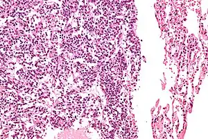 | |
| Micrograph of a typical pulmonary carcinoid tumour. | |
| Specialty | Oncology |
Typical pulmonary carcinoid tumour is a subtype of pulmonary carcinoid tumour. It is an uncommon low-grade malignant lung mass that is most often in the central airways of the lung.[1]
Signs and symptoms
Lung carcinoids typically present with a cough or hemoptysis.[2] Findings may closely mimic malignant tumours of the lung, i.e. lung cancer.
Diagnosis
.jpg.webp)
The definitive diagnosis is rendered by a microscopic examination, after excision. Typical carcinoids have cells with stippled chromatin and a moderate quantity of cytoplasm. They typically have few mitoses and lack necrosis. By definition, they are greater than 4 mm in largest dimension; smaller lesions are referred to as pulmonary carcinoid tumourlets.
The differential diagnosis of typical pulmonary carcinoid tumour includes: atypical pulmonary carcinoid tumour, pulmonary carcinoid tumourlet and lung adenocarcinoma.
 Very high magnification
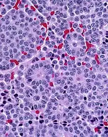 With prominent rosettes
Treatment
Typical carcinoids are usually treated with surgical excision.
See also
References
- ↑ Meisinger QC, Klein JS, Butnor KJ, Gentchos G, Leavitt BJ (November 2011). "CT features of peripheral pulmonary carcinoid tumors". AJR. American Journal of Roentgenology. 197 (5): 1073–1080. doi:10.2214/AJR.10.5954. PMID 22021498.
- ↑ Gungor S, Damadoglu E, Aybatli A, Yilmaz A, Kir A, Akkaya E (July 2006). "Typical pulmonary carcinoid tumors: presentation and outcome of 24 cases". Medical Science Monitor. 12 (7): CR315–CR318. PMID 16810137.