ERCC6
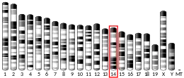
DNA excision repair protein ERCC-6 (also CS-B protein) is a protein that in humans is encoded by the ERCC6 gene.[4][5][6] The ERCC6 gene is located on the long arm of chromosome 10 at position 11.23.[7]
Having 1 or more copies of a mutated ERCC6 causes Cockayne syndrome, type II.
Function
DNA can be damaged by ultraviolet radiation, toxins, radioactive substances, and reactive biochemical intermediates like free radicals. The ERCC6 protein is involved in repairing the genome when specific genes undergoing transcription (dubbed active genes) are inoperative; as such, ERCC6 serves as a transcription-coupled excision repair protein, being one of the fundamental enzymes in active gene repair.[7]
Structure and Mechanism
CSB has been found to exhibit ATPase properties; there are contradictory publications regarding the effect of ATP concentration on CSB's activity.[8] The most recent evidence suggests that ADP/AMP allosterically regulate CSB.[6] As such, it has been speculated that CSB may promote protein complex formation at repair sites subject to the ATP to ADP charge ratio.
Conservation of helicase motifs in eukaryote CSB is evident; all seven major domains of the protein are conserved among numerous RNA and DNA helicases. Detailed structural analysis of CSB has been performed; motifs I, Ia, II, and III are collectively called domain 1, while motifs IV, V, and VI comprise domain 2. These domains wrap around an interdomain cleft involved in ATP binding and hydrolysis. Motifs III and IV are in close proximity to the active site; hence, residues in these regions stabilize ATP/ADP binding via hydrogen bonding.[9] Domain 2 has been proposed to affect DNA binding after induced conformational changes stemming from ATP hydrolysis. Specific residues involved in gene binding have yet to be identified.[10]
The evolutionary roots of CSB has led some to contend that it exhibits helicase activity.[11] Evidence for the helicase properties of CSB is highly disputed; yet, it has been found the protein participates in intracellular trafficking, a traditional role of helicases. The complex interactions between DNA repair proteins suggest that eukaryote CSB upholds some but not all of the functions of its prokaryotic precursors.[12]
Interactions
CSB has been shown to interact with P53.[13][14]
CSB has been shown to act as chromatin remodeling factor for RNA Polymerase II. When RNA Polymerase II is stalled by a mistake in the genome, CSB remodels the DNA double helix so as to allow access by repair enzymes to the lesion.[15]
CSB is involved in the base excision repair (BER) pathway. This is due to demonstrated interactions with human AP endonuclease, though interactions between recombinant CSB and E. coli endonuclease IV as well as human N-terminus AP endonuclease fragments have not been observed in vitro. Specifically, CSB stimulates the AP site incision activity of AP endonuclease independent of ATP.[16]
In addition to the BER pathway, CSB is heavily integrated in the nucleotide excision repair (NER) pathway. While BER utilizes glycosylases to recognize and correct non-bulky lesions, NER is particularly versatile in repairing DNA damaged by UV radiation via the removal of oxidized bases. CSB's role in NER is best manifested by interactions with T cell receptors, in which protein collaboration is key in effective antigen binding.[17]
Neurogenesis and Neural Differentiation
ERCC6 knockout within human neural progenitor cells has been shown to decrease both neurogenesis and neural differentiation. Both mechanisms are key in brain development, explaining characteristic cognitive deficiencies of Cockayne syndrome - such as stunted development of the nervous system - that otherwise do not seem related to symptoms like photosensitivity and hearing loss.[18]
Cockayne syndrome
In humans, Cockayne syndrome (CS) is a rare autosomal recessive leukodystrophy (associated with the degradation of white matter). CS arises from germ line mutations in either of two genes, CSA(ERCC8) or CSB(ERCC6). About two thirds of CS patients have a mutation in the CSB(ERCC6) gene.[19] Mutations in ERCC6 that lead to CS deal with both the size of the protein as well as the specific amino acid residues utilized in biosynthesis. Patients exhibiting type II CS often have shortened and/or misfolded CSB that disrupt gene expression and transcription. The characteristic biological effect of malfunctioning ERCC6 is nerve cell death, resulting in premature aging and growth defects.[7]
The extent to which malfunctioning CSB hinders oxidative repair heavily influences patients' neurological functioning. The two subforms of the disorder (the latter of which corresponds to ERCC6 defects) - CS-A and CS-B - both cause problems in the oxidative repair, though CS-B patients more often exhibit nerve system problems stemming from damage to this pathway. Most type II CS patients exhibit photosensitivity as per the heavily oxidative properties of UV light.[20][21]
While two copies of mutated ERCC6 result in CS, possession of a single copy of mutated ERCC6 gene is associated with similar but milder defects as CS, including retinal dystrophy, cardiac arrhythmias, and immunodeficiency.[22] Individuals who are heterozygote carriers are therefore at increased risk of similar pleiotropic disorders as homozygote carriers afflicted with CS.
DNA repair
CSB and CSA proteins are considered to function in transcription coupled nucleotide excision repair (TC-NER). CSB and CSA deficient cells are unable to preferentially repair UV-induced cyclobutane pyrimidine dimers in actively transcribed genes, consistent with a failed TC-NER response.[23] CSB also accumulates at sites of DNA double-strand breaks in a transcription dependent manner and influences double-strand break repair.[24] CSB protein facilitates homologous recombinational repair of double-strand breaks and represses non-homologous end joining.[24]
In a damaged cell, the CSB protein localizes to sites of DNA damage. CSB recruitment to damaged sites is influenced by the type of DNA damage and is, most rapid and robust as follows: interstrand crosslinks > double-strand breaks > monoadducts > oxidative damages.[19] The CSB protein interacts with SNM1A(DCLRE1A) protein, a 5’- 3’ exonuclease, to promote the removal of DNA interstrand crosslinks.[25]
Implications in cancer
Single-nucleotide polymorphisms in the ERCC6 gene have been correlated with significantly increased risk of certain forms of cancer. A specific mutation at the 1097 position (M1097V) as well as polymorphisms at amino acid residue 1413 have been associated with heightened risk of bladder cancer for experimental subjects in Taiwan; moreover, M1097V has been argued to play a key role in pathogenesis.[26] Rs1917799 polymorphism has been associated with increased risk of gastric cancer for Chinese experimental subjects,[27] and mutations at codon 399 have been correlated to the onset of oral cancers among Taiwanese patients.[28] Another study found a diverse set of mutations in the ERCC6 gene among Chinese lung cancer patients versus the general population (in terms of statistical significance), but failed to identify specific polymorphisms correlated with the patients' illness.[29]
Faulty DNA repair is implicated causally in tumor development due to malfunctioning proteins' inability to correct genes responsible for apoptosis and cell growth. Yet, the vast majority of studies regarding the effects of ERCC6 knockout or mutations on cancer are based upon statistical correlations of available patient data as opposed to mechanistic analysis of in vivo cancer onset. Hence, confounding based on protein-protein, protein-substrate, and/or substrate-substrate interactions disallows conclusions positing mutations in ERCC6 cause cancer on an individual basis.
References
- GRCm38: Ensembl release 89: ENSMUSG00000054051 - Ensembl, May 2017
- "Human PubMed Reference:". National Center for Biotechnology Information, U.S. National Library of Medicine.
- "Mouse PubMed Reference:". National Center for Biotechnology Information, U.S. National Library of Medicine.
- Troelstra C, van Gool A, de Wit J, Vermeulen W, Bootsma D, Hoeijmakers JH (Dec 1992). "ERCC6, a member of a subfamily of putative helicases, is involved in Cockayne's syndrome and preferential repair of active genes". Cell. 71 (6): 939–53. doi:10.1016/0092-8674(92)90390-X. hdl:1765/3041. PMID 1339317. S2CID 30671008.
- Muftuoglu M, de Souza-Pinto NC, Dogan A, Aamann M, Stevnsner T, Rybanska I, Kirkali G, Dizdaroglu M, Bohr VA (Apr 2009). "Cockayne syndrome group B protein stimulates repair of formamidopyrimidines by NEIL1 DNA glycosylase". The Journal of Biological Chemistry. 284 (14): 9270–9. doi:10.1074/jbc.M807006200. PMC 2666579. PMID 19179336.
- "Entrez Gene: ERCC6 excision repair cross-complementing rodent repair deficiency, complementation group 6".
- NIH. "ERCC6 Gene." Genetics Home Reference. National Institutes of Health, 16 Feb. 2015. Web. 22 Feb. 2015. <http://ghr.nlm.nih.gov/gene/ERCC6>.
- Selby CP, Sancar A (Jan 17, 1997). "Human transcription-repair coupling factor CSB/CSB is a DNA-stimulated ATPase but is not a helicase and does not disrupt the ternary transcription complex of stalled RNA polymerase II". J Biol Chem. 272 (3): 1885–90. doi:10.1074/jbc.272.3.1885. PMID 8999876.
- Durr H, Korner C, Muller M, Hickmann V, Hopfner KP (2005). "X-ray structures of the Sulfolobus solfataricus SWI2/SNF2 ATPase core and its complex with DNA". Cell. 121 (3): 363–373. doi:10.1016/j.cell.2005.03.026. PMID 15882619.
- Lewis R, Durr H, Hopfner KP, Michaelis J (2008). "Conformational changes of a Swi2/ Snf2 ATPase during its mechano-chemical cycle". Nucleic Acids Res. 36 (6): 1881–1890. doi:10.1093/nar/gkn040. PMC 2346605. PMID 18267970.
- Troelstra C, van Gool A, de Wit J, Vermeulen W, Bootsma D, Hoeijmakers JH (1993). "CSB, a member of a subfamily of putative helicases, is involved in Cockayne's syndrome and preferential repair of active genes". Cell. 71 (6): 939–53. doi:10.1016/0092-8674(92)90390-x. hdl:1765/3041. PMID 1339317. S2CID 30671008.
- Boulikas, T (March–April 1997). "Nuclear import of DNA repair proteins". Anticancer Research. 17 (2A): 843–63. PMID 9137418.
- Wang XW, Yeh H, Schaeffer L, Roy R, Moncollin V, Egly JM, Wang Z, Freidberg EC, Evans MK, Taffe BG (Jun 1995). "p53 modulation of TFIIH-associated nucleotide excision repair activity". Nature Genetics. 10 (2): 188–95. doi:10.1038/ng0695-188. hdl:1765/54884. PMID 7663514. S2CID 38325851.
- Yu A, Fan HY, Liao D, Bailey AD, Weiner AM (May 2000). "Activation of p53 or loss of the Cockayne syndrome group B repair protein causes metaphase fragility of human U1, U2, and 5S genes". Molecular Cell. 5 (5): 801–10. doi:10.1016/S1097-2765(00)80320-2. PMID 10882116.
- Newman JC, Bailey AD, Weiner AM (Jun 2006). "Cockayne syndrome group B protein (CSB) plays a general role in chromatin maintenance and remodeling". Proceedings of the National Academy of Sciences of the United States of America. 103 (25): 9313–8. Bibcode:2006PNAS..103.9613N. doi:10.1073/pnas.0510909103. PMC 1480455. PMID 16772382.
- Wong HK, Muftuoglu M, Beck G, Imam SZ, Bohr VA, Wilson DM (June 2007). "Cockayne syndrome B protein stimulates apurinic endonuclease 1 activity and protects against agents that introduce base excision repair intermediates". Nucleic Acids Research. 35 (12): 4103–13. doi:10.1093/nar/gkm404. PMC 1919475. PMID 17567611.
- Frosina G (Jul 2007). "The current evidence for defective repair of oxidatively damaged DNA in Cockayne syndrome". Free Radical Biology & Medicine. 43 (2): 165–77. doi:10.1016/j.freeradbiomed.2007.04.001. PMID 17603927.
- Ciaffardini F, Nicolai S, Caputo M, Canu G, Paccosi E, Costantino M, Frontini M, Balajee AS, Proietti-De-Santis L (2014). "The cockayne syndrome B protein is essential for neuronal differentiation and neuritogenesis". Cell Death Dis. 5 (5): e1268. doi:10.1038/cddis.2014.228. PMC 4047889. PMID 24874740.
- Iyama T, Wilson DM (2016). "Elements That Regulate the DNA Damage Response of Proteins Defective in Cockayne Syndrome". J. Mol. Biol. 428 (1): 62–78. doi:10.1016/j.jmb.2015.11.020. PMC 4738086. PMID 26616585.
- Laugel, V., C. Dalloz, M. Durrand, and H. Dollfus. "Mutation Update for the CSB/ERCC6 and CSA/ERCC8 Genes Involved in Cockayne Syndrome." Human Mutation. Human Genome Variation Society, 5 Nov. 2009. Web. 22 Feb. 2015. <http://onlinelibrary.wiley.com/doi/10.1002/humu.21154/epdf>.
- Nardo T, Oneda R, Spivak G, Mortier L, Thomas P, Orioli D, Laugel V, Stary A, Hanawalt PC, Sarasin A, Stefanini M (2009). "A UV-sensitive syndrome patient with a specific CSA mutation reveals separable roles for CSA in response to UV and oxidative DNA damage". Proc Natl Acad Sci USA. 106 (15): 6209–6214. Bibcode:2009PNAS..106.6209N. doi:10.1073/pnas.0902113106. PMC 2667150. PMID 19329487.
- Forrest, Iain S.; Chaudhary, Kumardeep; Vy, Ha My T.; Bafna, Shantanu; Kim, Soyeon; Won, Hong-Hee; Loos, Ruth J. F.; Cho, Judy; Pasquale, Louis R.; Nadkarni, Girish N.; Rocheleau, Ghislain (2021). "Genetic pleiotropy of ERCC6 loss-of-function and deleterious missense variants links retinal dystrophy, arrhythmia, and immunodeficiency in diverse ancestries". Human Mutation. 42 (8): 969–977. doi:10.1002/humu.24220. ISSN 1098-1004. PMC 8295228. PMID 34005834.
- van Hoffen A, Natarajan AT, Mayne LV, van Zeeland AA, Mullenders LH, Venema J (1993). "Deficient repair of the transcribed strand of active genes in Cockayne's syndrome cells". Nucleic Acids Res. 21 (25): 5890–5. doi:10.1093/nar/21.25.5890. PMC 310470. PMID 8290349.
- Batenburg NL, Thompson EL, Hendrickson EA, Zhu XD (2015). "Cockayne syndrome group B protein regulates DNA double-strand break repair and checkpoint activation". EMBO J. 34 (10): 1399–416. doi:10.15252/embj.201490041. PMC 4491999. PMID 25820262.
- Iyama T, Lee SY, Berquist BR, Gileadi O, Bohr VA, Seidman MM, McHugh PJ, Wilson DM (2015). "CSB interacts with SNM1A and promotes DNA interstrand crosslink processing". Nucleic Acids Res. 43 (1): 247–58. doi:10.1093/nar/gku1279. PMC 4288174. PMID 25505141.
- Chang CH, Chiu CF, Wang HC, Wu HC, Tsai RY, Tsai CW, Wang RF, Wang CH, Tsou YA, Bau DT (2009). "Significant association of ERCC6 single nucleotide polymorphisms with bladder cancer susceptibility in Taiwan". Anticancer Res. 29 (12): 5121–4. PMID 20044625.
- Liu JW, He CY, Sun LP, Xu Q, Xing CZ, Yuan Y (2013). "The DNA repair gene ERCC6 rs1917799 polymorphism is associated with gastric cancer risk in Chinese". Asian Pac. J. Cancer Prev. 14 (10): 6103–8. doi:10.7314/apjcp.2013.14.10.6103. PMID 24289633.
- Chiu CF, Tsai MH, Tseng HC, Wang CL, Tsai FJ, Lin CC, Bau DT (2008). "A novel single nucleotide polymorphism in ERCC6 gene is associated with oral cancer susceptibility in Taiwanese patients". Oral Oncol. 44 (6): 582–6. doi:10.1016/j.oraloncology.2007.07.006. PMID 17933579.
- Ma H, Hu Z, Wang H, Jin G, Wang Y, Sun W, Chen D, Tian T, Jin L, Wei Q, Lu D, Huang W, Shen H (2009). "ERCC6/CSB gene polymorphisms and lung cancer risk". Cancer Lett. 273 (1): 172–6. doi:10.1016/j.canlet.2008.08.002. PMID 18789574.
Further reading
- Cleaver JE, Thompson LH, Richardson AS, States JC (1999). "A summary of mutations in the UV-sensitive disorders: xeroderma pigmentosum, Cockayne syndrome, and trichothiodystrophy". Human Mutation. 14 (1): 9–22. doi:10.1002/(SICI)1098-1004(1999)14:1<9::AID-HUMU2>3.0.CO;2-6. PMID 10447254. S2CID 24148589.
- Troelstra C, Landsvater RM, Wiegant J, van der Ploeg M, Viel G, Buys CH, Hoeijmakers JH (Apr 1992). "Localization of the nucleotide excision repair gene ERCC6 to human chromosome 10q11-q21". Genomics. 12 (4): 745–9. doi:10.1016/0888-7543(92)90304-B. hdl:1765/3037. PMID 1349298.
- Fryns JP, Bulcke J, Verdu P, Carton H, Kleczkowska A, Van den Berghe H (Sep 1991). "Apparent late-onset Cockayne syndrome and interstitial deletion of the long arm of chromosome 10 (del(10)(q11.23q21.2))". American Journal of Medical Genetics. 40 (3): 343–4. doi:10.1002/ajmg.1320400320. PMID 1951442.
- Troelstra C, Odijk H, de Wit J, Westerveld A, Thompson LH, Bootsma D, Hoeijmakers JH (Nov 1990). "Molecular cloning of the human DNA excision repair gene ERCC-6". Molecular and Cellular Biology. 10 (11): 5806–13. doi:10.1128/MCB.10.11.5806. PMC 361360. PMID 2172786.
- Wang XW, Yeh H, Schaeffer L, Roy R, Moncollin V, Egly JM, Wang Z, Freidberg EC, Evans MK, Taffe BG (Jun 1995). "p53 modulation of TFIIH-associated nucleotide excision repair activity". Nature Genetics. 10 (2): 188–95. doi:10.1038/ng0695-188. hdl:1765/54884. PMID 7663514. S2CID 38325851.
- Henning KA, Li L, Iyer N, McDaniel LD, Reagan MS, Legerski R, Schultz RA, Stefanini M, Lehmann AR, Mayne LV, Friedberg EC (Aug 1995). "The Cockayne syndrome group A gene encodes a WD repeat protein that interacts with CSB protein and a subunit of RNA polymerase II TFIIH". Cell. 82 (4): 555–64. doi:10.1016/0092-8674(95)90028-4. PMID 7664335.
- Troelstra C, Hesen W, Bootsma D, Hoeijmakers JH (Feb 1993). "Structure and expression of the excision repair gene ERCC6, involved in the human disorder Cockayne's syndrome group B". Nucleic Acids Research. 21 (3): 419–26. doi:10.1093/nar/21.3.419. PMC 309134. PMID 8382798.
- Iyer N, Reagan MS, Wu KJ, Canagarajah B, Friedberg EC (Feb 1996). "Interactions involving the human RNA polymerase II transcription/nucleotide excision repair complex TFIIH, the nucleotide excision repair protein XPG, and Cockayne syndrome group B (CSB) protein". Biochemistry. 35 (7): 2157–67. doi:10.1021/bi9524124. PMID 8652557. S2CID 21846012.
- Selby CP, Sancar A (Jan 1997). "Human transcription-repair coupling factor CSB/ERCC6 is a DNA-stimulated ATPase but is not a helicase and does not disrupt the ternary transcription complex of stalled RNA polymerase II". The Journal of Biological Chemistry. 272 (3): 1885–90. doi:10.1074/jbc.272.3.1885. PMID 8999876.
- Boulikas T (1997). "Nuclear import of DNA repair proteins". Anticancer Research. 17 (2A): 843–63. PMID 9137418.
- Selby CP, Sancar A (Oct 1997). "Cockayne syndrome group B protein enhances elongation by RNA polymerase II". Proceedings of the National Academy of Sciences of the United States of America. 94 (21): 11205–9. Bibcode:1997PNAS...9411205S. doi:10.1073/pnas.94.21.11205. PMC 23417. PMID 9326587.
- Tantin D, Kansal A, Carey M (Dec 1997). "Recruitment of the putative transcription-repair coupling factor CSB/ERCC6 to RNA polymerase II elongation complexes". Molecular and Cellular Biology. 17 (12): 6803–14. doi:10.1128/MCB.17.12.6803. PMC 232536. PMID 9372911.
- Mallery DL, Tanganelli B, Colella S, Steingrimsdottir H, van Gool AJ, Troelstra C, Stefanini M, Lehmann AR (Jan 1998). "Molecular analysis of mutations in the CSB (ERCC6) gene in patients with Cockayne syndrome". American Journal of Human Genetics. 62 (1): 77–85. doi:10.1086/301686. PMC 1376810. PMID 9443879.
- Lindsay HD, Griffiths DJ, Edwards RJ, Christensen PU, Murray JM, Osman F, Walworth N, Carr AM (Feb 1998). "S-phase-specific activation of Cds1 kinase defines a subpathway of the checkpoint response in Schizosaccharomyces pombe". Genes & Development. 12 (3): 382–95. doi:10.1101/gad.12.3.382. PMC 316487. PMID 9450932.
- Tantin D (Oct 1998). "RNA polymerase II elongation complexes containing the Cockayne syndrome group B protein interact with a molecular complex containing the transcription factor IIH components xeroderma pigmentosum B and p62". The Journal of Biological Chemistry. 273 (43): 27794–9. doi:10.1074/jbc.273.43.27794. PMID 9774388.
- Dianov G, Bischoff C, Sunesen M, Bohr VA (Mar 1999). "Repair of 8-oxoguanine in DNA is deficient in Cockayne syndrome group B cells". Nucleic Acids Research. 27 (5): 1365–8. doi:10.1093/nar/27.5.1365. PMC 148325. PMID 9973627.
- Colella S, Nardo T, Mallery D, Borrone C, Ricci R, Ruffa G, Lehmann AR, Stefanini M (May 1999). "Alterations in the CSB gene in three Italian patients with the severe form of Cockayne syndrome (CS) but without clinical photosensitivity". Human Molecular Genetics. 8 (5): 935–41. doi:10.1093/hmg/8.5.935. PMID 10196384.
- Cheng L, Guan Y, Li L, Legerski RJ, Einspahr J, Bangert J, Alberts DS, Wei Q (Sep 1999). "Expression in normal human tissues of five nucleotide excision repair genes measured simultaneously by multiplex reverse transcription-polymerase chain reaction". Cancer Epidemiology, Biomarkers & Prevention. 8 (9): 801–7. PMID 10498399.
External links
- GeneReviews/NCBI/NIH/UW entry on Cockayne syndrome
- Overview of all the structural information available in the PDB for UniProt: Q03468 (DNA excision repair protein ERCC-6) at the PDBe-KB.


