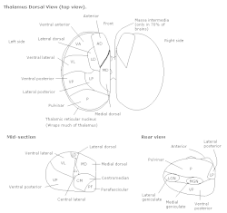
Centromedian nucleus
In the anatomy of the brain, the centromedian nucleus, also known as the centrum medianum, (CM or Cm-Pf) is a part of the intralaminar thalamic nuclei (ITN) in the thalamus. There are two centromedian nuclei arranged bilaterally.
| Centromedian nucleus | |
|---|---|
| Details | |
| Part of | Intralaminar thalamic nuclei |
| Identifiers | |
| Latin | nucleus centromedianus thalami |
| Acronym(s) | CM or Cm-Pf |
| NeuroNames | 323 |
| NeuroLex ID | birnlex_805 |
| TA98 | A14.1.08.618 |
| FMA | 62165 |
| Anatomical terms of neuroanatomy | |
In humans, it contains about 2000 neurons per cubic millimetre and has a volume of about 310 cubic millimetres with 664,000 neurons in total.[1]
Input and output
It sends nerve fibres to the subthalamic nucleus and putamen.[2] It receives nerve fibres from the cerebral cortex, vestibular nuclei, globus pallidus, superior colliculus, reticular formation, and spinothalamic tract.
Function
Its physiological role involves attention and arousal, including control of the level of cortical activity. Some frequencies of extracellular electrical stimulation of the centromedian nucleus can cause absence seizures (temporary loss of consciousness) although electrical stimulation can be of therapeutic use in intractable epilepsy and Tourette's syndrome. Specifically, centromedian nucleus has been proposed to be a target for neuromodulation-based treatment of generalized epilepsy.[3] General anaesthetics specifically suppress activity in the ILN, including the centromedian nucleus. Complete bilateral lesions of the centromedian nucleus can lead to states normally associated with brain death such as coma, death, persistent vegetative state, forms of mutism and severe delirium. Unilateral lesions can lead to unilateral thalamic neglect.
A patient with electrodes implanted into more than 50 different regions in his brain (including regions giving him orgasmic feelings) choose to self stimulate the electrode in his centromedian nucleus more than all other electrodes. The patients explanation of this: "The subject reported that he was almost able to recall a memory during this stimulation, but he could not quite grasp it. The frequent selfstimulations were an endeavor to bring this elusive memory into clear focus."[4]
Additional images
 Thalamus
 Thalamus
Notes and references
- Henderson J, Carpenter K, Cartwright H, Halliday G (2000). "Loss of thalamic intralaminar nuclei in progressive supranuclear palsy and Parkinson's disease: clinical and therapeutic implications". Brain. 123 ( Pt 7) (7): 1410–1421. doi:10.1093/brain/123.7.1410. PMID 10869053. Archived from the original on 2004-12-25. Retrieved 2004-09-24.
- Powell, T. P. S.; Cowan W. M. (1967). "The interpretation of the degenerative changes in the intralaminar nuclei of the thalamus". Journal of Neurology, Neurosurgery & Psychiatry. 30 (2): 140–153. doi:10.1136/jnnp.30.2.140. PMC 496153. PMID 4962197.
- Kokkinos, Vasileios; Urban, Alexandra; Sisterson, Nathaniel D.; Li, Ningfei; Corson, Danielle; Richardson, R Mark (2020). "Responsive Neurostimulation of the Thalamus Improves Seizure Control in Idiopathic Generalized Epilepsy: A Case Report". Neurosurgery. pp. E578–E583. doi:10.1093/neuros/nyaa001. PMID 32023343.
- "Electrical Self-Stimulation of the Brain in 4 1". 1963. CiteSeerX 10.1.1.188.7109.
{{cite journal}}: Cite journal requires|journal=(help)
External links
- Illustration and text: coro97/contents.htm at the University of Wisconsin-Madison Medical school - The Global Thalamus (See level 12, the centromedian nucleus is labelled CM)
- Schiff N, Plum F (2000). "The role of arousal and "gating" systems in the neurology of impaired consciousness". Journal of Clinical Neurophysiology. 17 (5): 438–52. doi:10.1097/00004691-200009000-00002. PMID 11085547. S2CID 17983908.
- Joe Bogen's Consciousness Page (Professor Bogen) at California Institute of Technology
- NIF Search - Centromedian Nucleus via the Neuroscience Information Framework