Erythrocyte sedimentation rate
| Erythrocyte sedimentation rate | |
|---|---|
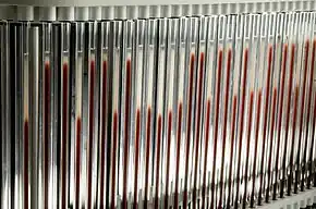
 Westergren pipet array on StaRRsed automated ESR analyzer. The ESR is the height (in mm) of the colourless portion at the top of the pipette after one hour. | |
| Synonyms | sedimentation rate, Westergren ESR, ESR, sed rate[1] |
| Reference range | Male: ≤ age/2 ; Female: ≤ (age + 10)/2.[2] (Unit: mm/hour).[2] |
| Purpose | Detection of inflammation in body.[1] |
| Test of | The rate of sedimentation of erythrocytes in a vertical tube over an hour.[1] |
| Based on | The millimeters of transparent fluid present at the top portion of the vertical tube after an hour.[1] |
| MeSH | D001799 |
| MedlinePlus | 003638 |
| LOINC | 30341-2 |
The erythrocyte sedimentation rate (ESR or sed rate) is the rate at which red blood cells in anticoagulated whole blood descend in a standardized tube over a period of one hour. It is a common hematology test, and is a non-specific measure of inflammation. To perform the test, anticoagulated blood is traditionally placed in an upright tube, known as a Westergren tube, and the distance which the red blood cells fall is measured and reported in mm at the end of one hour.[3]
Since the introduction of automated analyzers into the clinical laboratory, the ESR test has been automatically performed.
The ESR is governed by the balance between pro-sedimentation factors, mainly fibrinogen, and those factors resisting sedimentation, namely the negative charge of the erythrocytes (zeta potential). When an inflammatory process is present, the high proportion of fibrinogen in the blood causes red blood cells to stick to each other. The red cells form stacks called rouleaux which settle faster, due to their increased density. Rouleaux formation can also occur in association with some lymphoproliferative disorders in which one or more paraproteins are secreted in high amounts. While abnormal in humans, rouleaux formation can be a normal physiological finding in horses, cats, and pigs.
The ESR is increased in inflammation, pregnancy, anemia, autoimmune disorders (such as rheumatoid arthritis and lupus), infections, some kidney diseases and some cancers (such as lymphoma and multiple myeloma). The ESR is decreased in polycythemia, hyperviscosity, sickle cell anemia, leukemia, chronic fatigue syndrome,[4] low plasma protein (due to liver or kidney disease) and congestive heart failure. Although increases in immunoglobulins usually increase the ESR, very high levels can reduce it again due to hyperviscosity of the plasma.[5] This is especially likely with IgM-class paraproteins, and to a lesser extent, IgA-class. The basal ESR is slightly higher in females.[6]
Stages
Erythrocyte sedimentation rate (ESR) is the measure of ability of erythrocytes (red blood cell) to fall through the blood plasma and accumulate together at the base of container in one hour.[7]
There are three stages in erythrocyte sedimentation:[8]
- Rouleaux formation
- Sedimentation or settling stage
- Packing stage - 10 minutes (sedimentation slows and cells start to pack at the bottom of the tube)
In normal conditions, the red blood cells are negatively charged and therefore repel each other rather than stacking. ESR is also reduced by high blood viscosity, which slows the rate of fall.[7]
Causes of elevation
The rate of erythrocyte sedimentation is affected by both inflammatory and non-inflammatory conditions.
Inflammation
In inflammatory conditions, fibrinogen, other clotting proteins, and alpha globulin are positively charged, thus increasing the ESR.[9] ESR begins to rise at 24 to 48 hours after the onset of acute self-limited inflammation, decreases slowly as inflammation resolves, and can take weeks to months to return to normal levels. For ESR values more than 100 mm/hour, there is a 90% probability that an underlying cause would be found upon investigation.[9]
Non-inflammatory conditions
In non-inflammatory conditions, plasma albumin concentration, size, shape, and number of red blood cells, and the concentration of immunoglobulin can affect the ESR. Non-inflammatory conditions that can cause raised ESR include anemia, kidney failure, obesity, ageing, and female sex.[7] ESR is also higher in women during menstruation and pregnancy.[9] The value of ESR does not change whether dialysis is performed or not. Therefore, ESR is not a reliable measure of inflammation in those with kidney injuries as the ESR value is already elevated.[10]
Causes of reduction
An increased number of red blood cells (polycythemia) causes reduced ESR as blood viscosity increases. Hemoglobinopathy such as sickle-cell disease can have low ESR due to an improper shape of red blood cells that impairs stacking.
Medical uses
Diagnosis
ESR can sometimes be useful in diagnosing diseases, such as multiple myeloma, temporal arteritis, polymyalgia rheumatica, various auto-immune diseases, systemic lupus erythematosus, rheumatoid arthritis, inflammatory bowel disease[11] and chronic kidney diseases. In many of these cases, the ESR may exceed 100 mm/hour.[12]
It is commonly used for a differential diagnosis for Kawasaki's disease (from Takayasu's arteritis; which would have a markedly elevated ESR) and it may be increased in some chronic infective conditions like tuberculosis and infective endocarditis. It is also elevated in subacute thyroiditis also known as DeQuervain's.
In markedly increased ESR of over 100 mm/h, infection is the most common cause (33% of cases in an American study), followed by cancer (17%), kidney disease (17%) and noninfectious inflammatory disorders (14%).[13] Yet, in pneumonia the ESR stays under 100.[14]
The usefulness of the ESR in current practice has been questioned by some, as it is a relatively imprecise and non-specific test compared to other available diagnostic tests.[15]
Disease severity
It is a component of the PCDAI (pediatric Crohn's disease activity index), an index for assessment of the severity of inflammatory bowel disease in children.
Monitoring response to therapy
The clinical usefulness of ESR is limited to monitoring the response to therapy in certain inflammatory diseases such as temporal arteritis, polymyalgia rheumatica and rheumatoid arthritis. It can also be used as a crude measure of response in Hodgkin's lymphoma. Additionally, ESR levels are used to define one of the several possible adverse prognostic factors in the staging of Hodgkin's lymphoma.
Normal values
Note: mm/h. = millimeters per hour.
Westergren's original normal values (men 3 mm/h and women 7 mm/h)[16] made no allowance for a person's age. Later studies from 1967 confirmed that ESR values tend to rise with age and to be generally higher in women.[17] Values of the ESR also appear to be slightly higher in normal populations of African-Americans than Caucasians of both genders.[18] Values also appear to be higher in anemic individuals than non-anemic individuals.[19]
Adults
The widely used[20] rule calculating normal maximum ESR values in adults (98% confidence limit) is given by a formula devised in 1983 from a study of ≈1000 individuals over the age of 20:[21] The normal values of ESR in men is age (in years) divided by 2; for women, the normal value is age (in years) plus 10, divided by 2.[9]
Other studies confirm a dependence of ESR on age and gender, as seen in the following:
ESR reference ranges from a large 1996 study of 3,910 healthy adults (NB. these use 95% confidence intervals rather than the 98% intervals used in the study used to derive the formula above, and because of the skewness of the data, these values appear to be less than expected from the above formula):[22]
| Age | 20 | 55 | 90 |
|---|---|---|---|
| Men—5% exceed | 12 | 14 | 19 |
| Women—5% exceed | 18 | 21 | 23 |
Children
Normal values of ESR have been quoted as 1[23] to 2[24] mm/h at birth, rising to 4 mm/h 8 days after delivery,[24] and then to 17 mm/h by day 14.[23]
Typical normal ranges quoted are:[6]
- Newborn: 0 to 2 mm/h
- Neonatal to puberty: 3 to 13 mm/h, but other laboratories place an upper limit of 20.[25]
Relation to C-reactive protein
C-reactive protein (CRP) is an acute phase protein. Therefore, it is a better marker for acute phase reaction than ESR. While ESR and CRP generally together correlate with the degree of inflammation, this is not always the case and results may be discordant[9] in 12.5% of the cases.[7] Cases with raised CRP but normal ESR may demonstrate a combination of infection and some other tissue damage such as myocardial infarction, and venous thromboembolism. Such inflammation may not be enough to raise the level of ESR. Those with high ESR usually do not have demonstrable inflammation. However, in cases of low grade bacterial infections of bone and joints such as coagulase negative staphylococcus (CoNS), and systemic lupus erythematosus (SLE), ESR can be a good marker for the inflammatory process. This may be due to the production of Interferon type I that inhibits the CRP production in liver cells during SLE.[26] CRP is a better marker for other autoimmune diseases such as polymyalgia rheumatica, giant cell arteritis,[7] post-operative sepsis, and neonatal sepsis. ESR may be reduced in those who are taking statins and non-steroidal anti-inflammatory drugs (NSAIDs).[9]
| High ESR/Low CRP[9] | Low ESR/High CRP[9] |
|---|---|
| Systemic lupus erythematosis
Bone and joint infections Ischemic stroke Waldenstrom's macroglobulinemia Multiple myeloma IgG4 related disease Low serum albumin |
Urinary tract, GI, lung and bloodstream infections
Myocardial infarction Venous thromboembolic disease Rheumatoid arthritis Low serum albumin |
History
The test was invented in 1897 by the Polish pathologist Edmund Biernacki.[27][28] In some parts of the world the test continues to be referred to as Biernacki's Reaction (Polish: odczyn Biernackiego, OB).[29] In 1918, Dr Robert Fåhræus noted that ESR differed only during pregnancy. Therefore, he suggested that ESR could be used as an indicator of pregnancy. In 1921, Dr Alf Vilhelm Albertsson Westergren used ESR to measure the disease outcome of tuberculosis. He defined the measurement standards of ESR which is still being used today.[7] Robert Fåhræus and Alf Vilhelm Albertsson Westergren are eponymously remembered for the 'Fahraeus-Westergren test' (abbreviated as FW test; in the UK, usually termed Westergren test),[29] which uses sodium citrate-anti-coagulated specimens.[30]
Research
According to a study released in 2015, a stop gain mutation in HBB gene (p. Gln40stop) was shown to be associated with ESR values in Sardinian population. The red blood cell count, whose values are inversely related to ESR, is affected in carriers of this SNP. This mutation is almost exclusive of the inhabitants of Sardinia and is a common cause of beta thalassemia.[31]
According to a 2010 study, there is a reverse correlation between ESR and general intelligence (IQ) in Swedish males aged 18–20[32]
References
- 1 2 3 4 "Erythrocyte Sedimentation Rate (ESR)". Lab Tests Online. Retrieved 2019-12-23.
- 1 2 Miller, A; Green, M; Robinson, D (1983-01-22). "Simple rule for calculating normal erythrocyte sedimentation rate". British Medical Journal (Clinical Research Ed.). BMJ. 286 (6361): 266. doi:10.1136/bmj.286.6361.266. ISSN 0959-8138. PMC 1546487. PMID 6402065.
- ↑ "Erythrocyte Sedimentation Rate (ESR)". labtestsonline.org. Retrieved 2019-12-12.
- ↑ Saha, Amit K; Schmidt, Brendan R; Wilhelmy, Julie; Nguyen, Vy; Do, Justin; Suja, Vineeth C; Nemat-Gorgani, Mohsen; Ramasubramanian, Anand K; Davis, Ronald W (2018-11-21). "Erythrocyte Deformability As a Potential Biomarker for Chronic Fatigue Syndrome". Blood. 132 (Suppl 1): 4874. doi:10.1182/blood-2018-99-117260. ISSN 0006-4971. Retrieved 2019-06-19.
- ↑ Eastham, R. D (1954). "The Erythrocyte Sedimentation Rate and the Plasma Viscosity". Journal of Clinical Pathology. 7 (2): 164–167. doi:10.1136/jcp.7.2.164. PMC 1023757. PMID 13163203.
- 1 2 MedlinePlus Encyclopedia: ESR
- 1 2 3 4 5 6 Harrison, Michael (June 2015). "Erythrocyte sedimentation rate and C-reactive protein". Australian Prescriber. 38 (3): 93–4. doi:10.18773/austprescr.2015.034. PMC 4653962. PMID 26648629.
- ↑ "Erythrocyte sedimentation rate (ESR)" (PDF). National Institute of Open Schooling, India. Retrieved 8 April 2018.
Sedimentation occurs in three stages. In the first stage, the red cells form rouleaux. In the second stage, sinking of the aggregates occurs at a constant speed. In the final stage, the rate of sedimentation slows as the aggregated cells pack at the bottom of the tube.
- 1 2 3 4 5 6 7 8 Bray C, Bell LN, Liang H, Haykal R, Kaiksow F, Mazza JJ, Yale SH (December 2016). "Erythrocyte Sedimentation Rate and C-reactive Protein Measurements and Their Relevance in Clinical Medicine" (PDF). WMJ. 115 (6): 317–21. PMID 29094869.
- ↑ Arik N, Bedir A, Günaydin M, Adam B, Halefi I (October 2000). "Do erythrocyte sedimentation rate and C-reactive protein levels have diagnostic usefulness in patients with renal failure?". Nephron (Letter to the editor). 86 (2): 224. doi:10.1159/000045760. PMID 11015011. S2CID 5967575.
- ↑ Liu S, Ren J, Xia Q, Wu X, Han G, Ren H, Yan D, Wang G, Gu G, Li J (December 2013). "Preliminary case-control study to evaluate diagnostic values of C-reactive protein and erythrocyte sedimentation rate in differentiating active Crohn's disease from intestinal lymphoma, intestinal tuberculosis and Behcet's syndrome". The American Journal of the Medical Sciences. 346 (6): 467–72. doi:10.1097/MAJ.0b013e3182959a18. PMID 23689052. S2CID 5173681.
- ↑ "Sedimentation Rate". WebMD. 2006-06-16. Retrieved 2008-03-01.
- ↑ Raiten, Daniel J; Ashour, Fayrouz A Sakr; Ross, A Catharine; Meydani, Simin N; Dawson, Harry D; Stephensen, Charles B; Brabin, Bernard J; Suchdev, Parminder S; van Ommen, Ben (2015). "Inflammation and Nutritional Science for Programs/Policies and Interpretation of Research Evidence (INSPIRE)". The Journal of Nutrition. 145 (5): 1039S–1108S. doi:10.3945/jn.114.194571. ISSN 0022-3166. PMC 4448820. PMID 25833893.
-Which cites: Fincher, Ruth-Marie E. (1986). "Clinical Significance of Extreme Elevation of the Erythrocyte Sedimentation Rate". Archives of Internal Medicine. 146 (8): 1581–3. doi:10.1001/archinte.1986.00360200151024. ISSN 0003-9926. PMID 3729639. - ↑ Falk, G.; Fahey, T. (2008). "C-reactive protein and community-acquired pneumonia in ambulatory care: systematic review of diagnostic accuracy studies". Family Practice. 26 (1): 10–21. doi:10.1093/fampra/cmn095. ISSN 0263-2136. PMID 19074757.
- ↑ Jurado, Rafael L. (2001). "Why Shouldn't We Determine the Erythrocyte Sedimentation Rate?". Clinical Infectious Diseases. 33 (4): 548–549. doi:10.1086/322605. PMID 11462193. S2CID 7244484.
- ↑ Westergren A (March 1957). "Diagnostic tests: the erythrocyte sedimentation rate range and limitations of the technique". Triangle; the Sandoz Journal of Medical Science. 3 (1): 20–5. PMID 13455726.
- ↑ Böttiger LE, Svedberg CA (April 1967). "Normal erythrocyte sedimentation rate and age". British Medical Journal. 2 (5544): 85–7. doi:10.1136/bmj.2.5544.85. PMC 1841240. PMID 6020854.
- ↑ Gillum RF (January 1993). "A racial difference in erythrocyte sedimentation". Journal of the National Medical Association. 85 (1): 47–50. PMC 2571720. PMID 8426384.
- ↑ Kanfer EJ, Nicol BA (January 1997). "Haemoglobin concentration and erythrocyte sedimentation rate in primary care patients" (Scanned & PDF). Journal of the Royal Society of Medicine. 90 (1): 16–8. doi:10.1177/014107689709000106. PMC 1296109. PMID 9059375.
- ↑ "Reference range (ESR)". GPnotebook.
- ↑ Miller A, Green M, Robinson D (January 1983). "Simple rule for calculating normal erythrocyte sedimentation rate". British Medical Journal. 286 (6361): 266. doi:10.1136/bmj.286.6361.266. PMC 1546487. PMID 6402065.
- ↑ Wetteland P, Røger M, Solberg HE, Iversen OH (September 1996). "Population-based erythrocyte sedimentation rates in 3910 subjectively healthy Norwegian adults. A statistical study based on men and women from the Oslo area". Journal of Internal Medicine. 240 (3): 125–31. doi:10.1046/j.1365-2796.1996.30295851000.x. PMID 8862121. S2CID 10871066. - listing upper reference levels expected to be exceeded only by chance in 5% of subjects
- 1 2 Adler SM, Denton RL (June 1975). "The erythrocyte sedimentation rate in the newborn period". The Journal of Pediatrics. 86 (6): 942–8. doi:10.1016/S0022-3476(75)80233-2. PMID 1168702.
- 1 2 Ibsen KK, Nielsen M, Prag J, Hørlyk H, Vrang C, Korner B, Peitersen B (1980). "The value of the micromethod erythrocyte sedimentation rate in the diagnosis of infections in newborns". Scandinavian Journal of Infectious Diseases. Supplementum. Suppl 23: 143–5. PMID 6937959.
- ↑ Mack DR, Langton C, Markowitz J, LeLeiko N, Griffiths A, Bousvaros A, et al. (June 2007). "Laboratory values for children with newly diagnosed inflammatory bowel disease". Pediatrics. 119 (6): 1113–9. doi:10.1542/peds.2006-1865. PMID 17545378. S2CID 5558076. Lay summary – NEJM Journal Watch (June 13, 2007).
{{cite journal}}: Cite uses deprecated parameter|lay-source=(help) - ↑ Enocsson, Helena; Sjöwall, Christopher; Skogh, Thomas; Eloranta, Maija-Leena; Rönnblom, Lars; Wetterö, Jonas (December 2009). "Interferon-α mediates suppression of C-reactive protein: Explanation for muted C-reactive protein response in lupus flares?". Arthritis & Rheumatism. 60 (12): 3755–3760. doi:10.1002/art.25042.
- ↑ Iłowiecki, Maciej (1981). Dzieje nauki polskiej. Warszawa: Wydawnictwo „Interpress”. p. 195. ISBN 978-83-223-1876-8.
- ↑ Edmund Faustyn Biernacki and eponymously named Biernacki's test at Who Named It?
- 1 2 Robert (Robin) Sanno Fåhræus and Alf Vilhelm Albertsson Westergren who are eponymously named for the Fåhræus-Westergren test (aka Westergren test) at Who Named It?
- ↑ International Council for Standardization in Haematology (Expert Panel on Blood Rheology) (March 1993). "ICSH recommendations for measurement of erythrocyte sedimentation rate. International Council for Standardization in Haematology (Expert Panel on Blood Rheology)". Journal of Clinical Pathology. 46 (3): 198–203. doi:10.1136/jcp.46.3.198. PMC 501169. PMID 8463411.
- ↑ Sidore C, Busonero F, Maschio A, Porcu E, Naitza S, Zoledziewska M, et al. (November 2015). "Genome sequencing elucidates Sardinian genetic architecture and augments association analyses for lipid and blood inflammatory markers". Nature Genetics. 47 (11): 1272–1281. doi:10.1038/ng.3368. PMC 4627508. PMID 26366554.
- ↑ Karlsson, Håkan; Ahlborg, Björn; Dalman, Christina; Hemmingsson, Tomas (August 2010). "Association between erythrocyte sedimentation rate and IQ in Swedish males aged 18–20". Brain, Behavior, and Immunity. 24 (6): 868–873. doi:10.1016/j.bbi.2010.02.009. PMID 20226851. S2CID 7185302.
External links
- Mediscuss on ESR
- Brigden ML (October 1999). "Clinical utility of the erythrocyte sedimentation rate". American Family Physician. 60 (5): 1443–50. PMID 10524488.
- ESR at Lab Tests Online