Interleukin 19
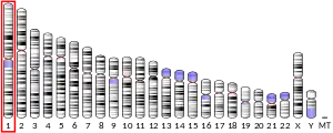

Interleukin 19 (IL-19) is a protein that belongs to the IL-10 cytokine subfamily. Human IL-19 is encoded by the IL-19 gene located on chromosome 1.[5]
Structure
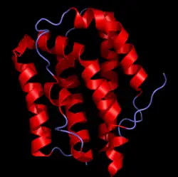
The IL-19 gene contains 7 exons. Secreted IL-19 protein is made from 159 amino acids in an alpha-helix structure.[5]
Function
IL-19 is an immunosuppressive cytokine that is responsible for both cell type-specific and system-specific down-regulation of inflammation. IL-19 interacts with both immune cells (macrophages, T cells, B cells) and non-immune cells (endothelial cells and brain resident glial cells, etc.).[6]
Induction of immune cells polarization
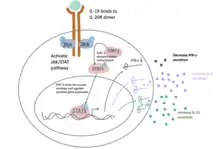
IL-19 is homologous to interleukin 20 (IL-20) and thus is able to bind the interleukin-20 receptor complex. IL-19 binding the dimer receptor IL20R leads to activation of the JAK/STAT signaling pathway. Following STAT protein entering the nucleus of a T-helper cell, the cell differentiates into a less inflammatory T-helper 2 cells through downregulation of interferon gamma (IFN-γ) and upregulation of interleukin 4(IL-4) and interleukin 10 (IL-10).[7][8][9]
Macrophages can also be polarized and have distinct functional phenotypes after receiving specific signals. When IL-19 binds to the IL-20R receptors on macrophages, it induces expression of IL-10, promoting the macrophage polarization from the inflammatory M1 type to an anti-inflammatory phenotype M2.[9] The IL-19 induced macrophage polarization reduces the pro-inflammatory cytokines produced by M1 macrophages, thus limiting the magnitude of the inflammatory response.
Protection against vascular inflammation
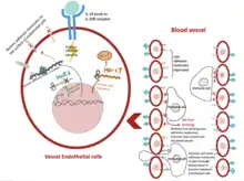
IL-19 is not detectable in healthy arteries, but frequently observed in damaged vessels. After receiving damage, IL-19 is secreted by vascular cells to recover from inflammation and protect tissue against further damage.[10]
Adhesion molecule regulation
IL-19 suppresses the expression of RNA-binding protein HuR, an mRNA stability factor.[11] HuR is responsible for stabilizing mRNA that codes for adhesion molecules excreted by activated macrophages, and later allow for neutrophil movement into peripheral or cardiac tissue. Through downregulation of this factor, fewer adhesion molecules are translated and expressed in the endothelial cell lined up in blood vessels. With fewer neutrophils entering cardiac tissue, IL-19 protects the vascular tissue from damage related to inflammatory processes.
Reduction of oxidative stress
IL-19 upregulates the expression of heme oxygenase-1 (HO-1) and reduces reactive oxygen species in human vascular smooth muscle cells.[12]
Immune cell polarization
The induction of anti-inflammatory cytokines (IL-10 and IL-4) and downregulating pro-inflammatory cytokines (IFN-γ) pushes a T helper cell away from T-helper 1 (Th1) to T-helper 2 (Th2).[12] During vascular infection, Th1 is the predominant T cells population and expresses interferon IFN-γ, tumor necrosis factor-α, and other pro-inflammatory cytokines, leading to vessel tissue damage[13] In contrast, Th2 cells secrete IL-4 and IL-10 and downregulate IFN-γ.[13] This dampens the inflammatory response by inhibiting pro-inflammatory gene expression. Additionally, macrophages receiving the IL-19 signal are polarized from M1 (pro-inflammatory) to M2 (anti-inflammatory).
Neuroprotection
The resident glial cells of the central nervous system participate in neuroinflammation initiation and regulation. Glial cells such as microglia and astrocytes produce inflammatory cytokines in response to foreign antigens and then produce immunosuppressive cytokines to resolve inflammation. IL-19, secreted by astrocytes, functions as an immunosuppressive signal in a manner of delayed kinetics of induction. The induction of IL-19 often occurs after immune response initiation as a secondary, anti-inflammatory, neuroprotective regulation to limit the inflammation.[14] Astrocytes produce IL-19 to interact with cells expressing IL-20 receptors (microglia etc.), in order to regulate cytokine secretion, which includes reducing the production of pro-inflammatory cytokines such as IL-6 and TNF-α.
IL-10 family
Interleukin-19 is a cytokine that belongs to the IL-10 family of cytokines along with several other interleukins including IL-10, IL-20, IL-22, IL-24, IL-26, and several virus-encoded cytokines. It signals through the same cell surface receptor (IL-20R) that is used by IL-20 and IL-24.
References
- 1 2 3 GRCh38: Ensembl release 89: ENSG00000142224 - Ensembl, May 2017
- 1 2 3 GRCm38: Ensembl release 89: ENSMUSG00000016524 - Ensembl, May 2017
- ↑ "Human PubMed Reference:". National Center for Biotechnology Information, U.S. National Library of Medicine.
- ↑ "Mouse PubMed Reference:". National Center for Biotechnology Information, U.S. National Library of Medicine.
- 1 2 "IL19 interleukin 19 [ Homo sapiens (human) ]".
{{cite web}}: CS1 maint: url-status (link) - ↑ Leigh T, Scalia RG, Autieri MV (September 2020). "Resolution of inflammation in immune and nonimmune cells by interleukin-19". American Journal of Physiology. Cell Physiology. 319 (3): C457–C464. doi:10.1152/ajpcell.00247.2020. PMC 7509264. PMID 32667867.
- ↑ Romagnani S (November 1999). "Th1/Th2 cells". Inflammatory Bowel Diseases. 5 (4): 285–94. doi:10.1097/00054725-199911000-00009. PMID 10579123.
- ↑ Gallagher G (October 2010). "Interleukin-19: multiple roles in immune regulation and disease". Cytokine & Growth Factor Reviews. 21 (5): 345–52. doi:10.1016/j.cytogfr.2010.08.005. PMID 20889366.
- 1 2 Azuma YT, Nakajima H, Takeuchi T (November 2011). "IL-19 as a potential therapeutic in autoimmune and inflammatory diseases". Current Pharmaceutical Design. 17 (34): 3776–80. doi:10.2174/138161211798357845. PMID 22103848.
- ↑ England RN, Autieri MV (2012). "Anti-inflammatory effects of interleukin-19 in vascular disease". International Journal of Inflammation. 2012: 253583. doi:10.1155/2012/253583. PMC 3403192. PMID 22844641.
- ↑ Scott DW, Patel RP (August 2013). "Targeting endothelial adhesion molecule mRNA to control inflammation: novel insights into potential anti-inflammatory effects of IL-19. Focus on "Interleukin-19 decreases leukocyte-endothelial cell interactions by reduction in endothelial cell adhesion molecule mRNA stability"". American Journal of Physiology. Cell Physiology. 305 (3): C253-4. doi:10.1152/ajpcell.00120.2013. PMID 23657567.
- 1 2 Autieri MV (2018). "IL-19 and Other IL-20 Family Member Cytokines in Vascular Inflammatory Diseases". Frontiers in Immunology. 9: 700. doi:10.3389/fimmu.2018.00700. PMC 5897441. PMID 29681905.
- 1 2 Lintermans LL, Stegeman CA, Heeringa P, Abdulahad WH (2014). "T cells in vascular inflammatory diseases". Frontiers in Immunology. 5: 504. doi:10.3389/fimmu.2014.00504. PMC 4196542. PMID 25352848.
- ↑ Burmeister AR, Marriott I (2018). "The Interleukin-10 Family of Cytokines and Their Role in the CNS". Frontiers in Cellular Neuroscience. 12: 458. doi:10.3389/fncel.2018.00458. PMC 6277801. PMID 30542269.
External links
- Overview of all the structural information available in the PDB for UniProt: Q9UHD0 (Interleukin-19) at the PDBe-KB.
This article incorporates text from the United States National Library of Medicine, which is in the public domain.