Kleeblattschaedel
| Kleeblattschaedel | |
|---|---|
| Other names | Cloverleaf skull, kleeblattschädel, isolated cloverleaf skull syndrome[1] |
 | |
| 1-day-old female infant with kleeblattschaedel | |
| Complications | Proptosis, recurrent corneal erosions, elbow ankylosis, hydrocephalus |
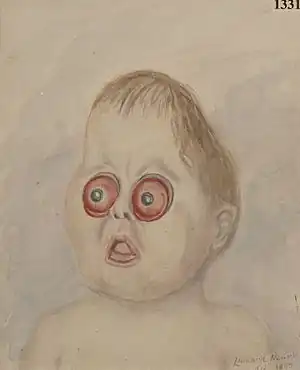
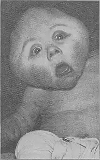
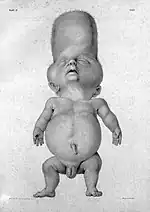
Kleeblattschaedel is a rare malformation of the head where there is a protrusion of the skull and broadening of the face.[2] This condition is a severe type of craniosynostosis.[3]
The condition can be both isolated or associated with other craniofacial dysostosises.[4] 85% of children with this condition have other anomalies.[5] Severe forms of the condition are often a sign of syndromic craniosynostosis combined with a grotesque constriction ring of the lambdoid structure and the squamosal bone or in another area.[4]
Name and etymology
Kleeblattschaedel (Kleeblattschädel) is German for "cloverleaf skull".[6] The disorder was named Kleeblattschaedel syndrome in 1958.[7] The German word is sometimes used in medical English, where it is often regarded as more or less naturalized, thus appearing in any combination of capitalized or not, with umlaut diacritic or not, and italicized or not.
History
The first case reported was back in 1849. The condition was first identified in 1960, and the first case in the United States was reported in 1965.[8]
Causes
The condition is caused by a premature fusing of the fibrous sutures.[9] The distinctive head shape seen in kleeblattschaedel is caused by the closure of the sagittal, coronal, and lambdoid sutures, with subsequent bulging of the cranial contents leading to a trilobate head shape.[7] The condition is also caused by absence of the coronal and lambdoid sutures.[10]
Conditions with kleeblattschaedel include:[11][12]
- Antley-Bixler syndrome
- Beare-Stevenson cutis gyrata syndrome
- Carpenter syndrome
- Cloverleaf skull-asphyxiating thoracic dysplasia syndrome[13]
- Cloverleaf skull-multiple congenital anomalies syndrome[14]
- Cranioectodermal dysplasia 2
- Crouzon syndrome
- Micromelic bone dysplasia with cloverleaf skull
- Mosaic trisomy 5[15]
- Muenke syndrome[16]
- Osteoglophonic dysplasia
- Pfeiffer syndrome
- Thanatophoric dysplasia, types 1 and 2
 Carpenter syndrome
 Crouzon syndrome
 Pfeiffer syndrome
 Thanatophoric dysplasia
Epidemiology
The condition occurs equally in both males as in females.[17]
References
- ↑ "Kleeblattschaedel syndrome | Genetic and Rare Diseases Information Center (GARD) – an NCATS Program". rarediseases.info.nih.gov. Retrieved 2021-09-03.
- ↑ Lindsey, Mary P. (2002-03-11). Dictionary of Mental Handicap. Routledge. p. 181. ISBN 978-1-134-97199-2.
- ↑ Wynbrandt, James; Ludman, Mark D. (2010-05-12). The Encyclopedia of Genetic Disorders and Birth Defects. Infobase Publishing. pp. 229–230. ISBN 978-1-4381-2095-9.
- 1 2 Kaiser, Georges L. (2012-12-13). Symptoms and Signs in Pediatric Surgery. Springer Science & Business Media. p. 79. ISBN 978-3-642-31161-1.
- ↑ Swaiman, Kenneth F.; Ashwal, Stephen M.; Ferriero, Donna M.; Schor, Nina F.; Finkel, Richard S.; Gropman, Andrea L.; Pearl, Phillip L.; Shevell, Michael (2017-09-21). Swaiman's Pediatric Neurology E-Book: Principles and Practice. Elsevier Health Sciences. pp. e582. ISBN 978-0-323-37481-1.
- ↑ Weaver, David D.; Brandt, Ira K. (1999). Catalog of Prenatally Diagnosed Conditions. JHU Press. p. 151. ISBN 978-0-8018-6044-7.
- 1 2 Radswiki, The. "Cloverleaf skull (craniosynostosis) | Radiology Reference Article | Radiopaedia.org". Radiopaedia. Retrieved 2023-01-28.
- ↑ Quinones-Hinojosa, Alfredo (2021-04-22). Schmidek and Sweet: Operative Neurosurgical Techniques E-Book: Indications, Methods and Results. Elsevier Health Sciences. p. 953. ISBN 978-0-323-41519-4.
- ↑ "Craniofacial Abnormalities". www.hopkinsmedicine.org. 8 August 2021. Retrieved 2021-10-02.
- ↑ Lewis, Mary (2017-07-26). Paleopathology of Children: Identification of Pathological Conditions in the Human Skeletal Remains of Non-Adults. Academic Press. p. 25. ISBN 978-0-12-410439-6.
- ↑ "Cloverleaf skull". Medgen - NCBI. Retrieved August 28, 2023.
- ↑ "Cloverleaf skull syndrome". Medgen - NCBI. Retrieved August 28, 2023.
- ↑ "Cloverleaf skull-asphyxiating thoracic dysplasia syndrome". Medgen - NCBI. Retrieved August 28, 2023.
- ↑ "Cloverleaf skull-multiple congenital anomalies syndrome". Medgen - NCBI. Retrieved August 28, 2023.
- ↑ "Mosaic trisomy 5". Medgen - NCBI. Retrieved August 28, 2023.
- ↑ "Muenke syndrome". Medgen - NCBI. Retrieved August 28, 2023.
- ↑ Ketonen, L. M.; Hiwatashi, A.; Sidhu, R.; Westesson, P.-L. (2005-12-05). Pediatric Brain and Spine: An Atlas of MRI and Spectroscopy. Springer Science & Business Media. p. 64. ISBN 978-3-540-26436-1.