Keratin 19
Keratin, type I cytoskeletal 19 also known as cytokeratin-19 (CK-19) or keratin-19 (K19) is a 40 kDa protein that in humans is encoded by the KRT19 gene.[5][6] Keratin 19 is a type I keratin.
| KRT19 | |||||||||||||||||||||||||||||||||||||||||||||||||||
|---|---|---|---|---|---|---|---|---|---|---|---|---|---|---|---|---|---|---|---|---|---|---|---|---|---|---|---|---|---|---|---|---|---|---|---|---|---|---|---|---|---|---|---|---|---|---|---|---|---|---|---|
| Identifiers | |||||||||||||||||||||||||||||||||||||||||||||||||||
| Aliases | KRT19, CK19, K19, K1CS, keratin 19 | ||||||||||||||||||||||||||||||||||||||||||||||||||
| External IDs | OMIM: 148020 MGI: 96693 HomoloGene: 1713 GeneCards: KRT19 | ||||||||||||||||||||||||||||||||||||||||||||||||||
| |||||||||||||||||||||||||||||||||||||||||||||||||||
| |||||||||||||||||||||||||||||||||||||||||||||||||||
| |||||||||||||||||||||||||||||||||||||||||||||||||||
| |||||||||||||||||||||||||||||||||||||||||||||||||||
| |||||||||||||||||||||||||||||||||||||||||||||||||||
| Wikidata | |||||||||||||||||||||||||||||||||||||||||||||||||||
| |||||||||||||||||||||||||||||||||||||||||||||||||||
Function
Keratin 19 is a member of the keratin family. The keratins are intermediate filament proteins responsible for the structural integrity of epithelial cells and are subdivided into cytokeratins and hair keratins.
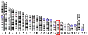
Keratin 19 is a type I keratin. The type I cytokeratins consist of acidic proteins which are arranged in pairs of heterotypic keratin chains. Unlike its related family members, this smallest known acidic cytokeratin is not paired with a basic cytokeratin in epithelial cells. It is specifically found in the periderm, the transiently superficial layer that envelops the developing epidermis. The type I cytokeratins are clustered in a region of chromosome 17q12-q21.[6]
Use as biomarker
KRT19 is also known as Cyfra 21-1.[7]
Due to its high sensitivity, KRT19 is the most used marker for the RT-PCR-mediated detection of tumor cells disseminated in lymph nodes, peripheral blood, and bone marrow of breast cancer patients. Depending on the assays, KRT19 has been shown to be both a specific and a non-specific marker. False positivity in such KRT19 RT-PCR studies include: illegitimate transcription (expression of small amounts of KRT19 mRNA by tissues in which it has no real physiological role), haematological disorders (KRT19 induction in peripheral blood cells by cytokines and growth factors, which circulate at higher concentrations in inflammatory conditions and neutropenia), the presence of pseudogenes (two KRT19 pseudogenes, KRT19a and KRT19b, have been identified, which have significant sequence homology to KRT19 mRNA. Subsequently, attempts to detect the expression of the authentic KRT19 may result in the detection of either or both of these pseudogenes), sample contamination (introduction of contaminating epithelial cells during peripheral blood sampling for subsequent RT-PCR analysis).[8] Moreover, Ck-19 is widely applied as post-operative diagnostic marker of papillary thyroid carcinoma.[9]
Keratin 19 is often used together with keratin 8 and keratin 18 to differentiate cells of epithelial origin from hematopoietic cells in tests that enumerate circulating tumor cells in blood.[10]
References
- GRCh38: Ensembl release 89: ENSG00000171345 - Ensembl, May 2017
- GRCm38: Ensembl release 89: ENSMUSG00000020911 - Ensembl, May 2017
- "Human PubMed Reference:". National Center for Biotechnology Information, U.S. National Library of Medicine.
- "Mouse PubMed Reference:". National Center for Biotechnology Information, U.S. National Library of Medicine.
- Schweizer J, Bowden PE, Coulombe PA, Langbein L, Lane EB, Magin TM, Maltais L, Omary MB, Parry DA, Rogers MA, Wright MW (July 2006). "New consensus nomenclature for mammalian keratins". The Journal of Cell Biology. 174 (2): 169–74. doi:10.1083/jcb.200603161. PMC 2064177. PMID 16831889.
- "Entrez Gene: KRT19 keratin 19".
- Nakata B, Takashima T, Ogawa Y, Ishikawa T, Hirakawa K (2004). "Serum CYFRA 21-1 (cytokeratin-19 fragments) is a useful tumour marker for detecting disease relapse and assessing treatment efficacy in breast cancer". Br J Cancer. 91 (5): 873–8. doi:10.1038/sj.bjc.6602074. PMC 2409884. PMID 15280913.
- Lacroix M (December 2006). "Significance, detection and markers of disseminated breast cancer cells". Endocrine-Related Cancer. 13 (4): 1033–67. doi:10.1677/ERC-06-0001. PMID 17158753.
- Dinets A, Pernemalm M, Kjellin H, Sviatoha V, Sofiadis A, Juhlin CC, Zedenius J, Larsson C, Lehtiö J, Höög A (May 2015). "Differential protein expression profiles of cyst fluid from papillary thyroid carcinoma and benign thyroid lesions". PLOS ONE. 10 (5): e0126472. Bibcode:2015PLoSO..1026472D. doi:10.1371/journal.pone.0126472. PMC 4433121. PMID 25978681.
- Allard WJ, Matera J, Miller MC, Repollet M, Connelly MC, Rao C, Tibbe AG, Uhr JW, Terstappen LW (October 2004). "Tumor cells circulate in the peripheral blood of all major carcinomas but not in healthy subjects or patients with nonmalignant diseases". Clinical Cancer Research. 10 (20): 6897–904. doi:10.1158/1078-0432.CCR-04-0378. PMID 15501967.
- Shi J, Sugrue SP (May 2000). "Dissection of protein linkage between keratins and pinin, a protein with dual location at desmosome-intermediate filament complex and in the nucleus". The Journal of Biological Chemistry. 275 (20): 14910–5. doi:10.1074/jbc.275.20.14910. PMID 10809736.
Further reading
- Otsuka Y, Ichikawa Y, Kunisaki C, Matsuda G, Akiyama H, Nomura M, Togo S, Hayashizaki Y, Shimada H (2007). "Correlating purity by microdissection with gene expression in gastric cancer tissue". Scandinavian Journal of Clinical and Laboratory Investigation. 67 (4): 367–79. doi:10.1080/00365510601046334. PMID 17558891. S2CID 36635804.
- Rasmussen HH, van Damme J, Puype M, Gesser B, Celis JE, Vandekerckhove J (December 1992). "Microsequences of 145 proteins recorded in the two-dimensional gel protein database of normal human epidermal keratinocytes". Electrophoresis. 13 (12): 960–9. doi:10.1002/elps.11501301199. PMID 1286667. S2CID 41855774.
- Bader BL, Magin TM, Hatzfeld M, Franke WW (August 1986). "Amino acid sequence and gene organization of cytokeratin no. 19, an exceptional tail-less intermediate filament protein". The EMBO Journal. 5 (8): 1865–75. doi:10.1002/j.1460-2075.1986.tb04438.x. PMC 1167052. PMID 2428612.
- Stasiak PC, Lane EB (December 1987). "Sequence of cDNA coding for human keratin 19". Nucleic Acids Research. 15 (23): 10058. doi:10.1093/nar/15.23.10058. PMC 306562. PMID 2447559.
- Eckert RL (February 1988). "Sequence of the human 40-kDa keratin reveals an unusual structure with very high sequence identity to the corresponding bovine keratin". Proceedings of the National Academy of Sciences of the United States of America. 85 (4): 1114–8. Bibcode:1988PNAS...85.1114E. doi:10.1073/pnas.85.4.1114. PMC 279716. PMID 2448790.
- Blumenberg M (1988). "Concerted gene duplications in the two keratin gene families". Journal of Molecular Evolution. 27 (3): 203–11. Bibcode:1988JMolE..27..203B. doi:10.1007/BF02100075. PMID 2458477. S2CID 5606956.
- Bader BL, Jahn L, Franke WW (December 1988). "Low level expression of cytokeratins 8, 18 and 19 in vascular smooth muscle cells of human umbilical cord and in cultured cells derived therefrom, with an analysis of the chromosomal locus containing the cytokeratin 19 gene". European Journal of Cell Biology. 47 (2): 300–19. PMID 2468493.
- Stasiak PC, Purkis PE, Leigh IM, Lane EB (May 1989). "Keratin 19: predicted amino acid sequence and broad tissue distribution suggest it evolved from keratinocyte keratins". The Journal of Investigative Dermatology. 92 (5): 707–16. doi:10.1111/1523-1747.ep12721500. PMID 2469734.
- Shezen E, Okon E, Ben-Hur H, Abramsky O (January 1995). "Cytokeratin expression in human thymus: immunohistochemical mapping". Cell and Tissue Research. 279 (1): 221–31. doi:10.1007/BF00300707. PMID 7534649. S2CID 23434233.
- Milisavljevic V, Freedberg IM, Blumenberg M (May 1996). "Close linkage of the two keratin gene clusters in the human genome". Genomics. 34 (1): 134–8. doi:10.1006/geno.1996.0252. PMID 8661035.
- Ceratto N, Dobkin C, Carter M, Jenkins E, Yao XL, Cassiman JJ, Aly MS, Bosco P, Leube R, Langbein L, Feo S, Romano V (1997). "Human type I cytokeratin genes are a compact cluster". Cytogenetics and Cell Genetics. 77 (3–4): 169–74. doi:10.1159/000134566. PMID 9284906.
- Kucharzik T, Lügering N, Schmid KW, Schmidt MA, Stoll R, Domschke W (January 1998). "Human intestinal M cells exhibit enterocyte-like intermediate filaments". Gut. 42 (1): 54–62. doi:10.1136/gut.42.1.54. PMC 1726964. PMID 9505886.
- Yang GP, Ross DT, Kuang WW, Brown PO, Weigel RJ (March 1999). "Combining SSH and cDNA microarrays for rapid identification of differentially expressed genes". Nucleic Acids Research. 27 (6): 1517–23. doi:10.1093/nar/27.6.1517. PMC 148347. PMID 10037815.
- Zhou X, Liao J, Hu L, Feng L, Omary MB (April 1999). "Characterization of the major physiologic phosphorylation site of human keratin 19 and its role in filament organization". The Journal of Biological Chemistry. 274 (18): 12861–6. doi:10.1074/jbc.274.18.12861. PMID 10212274.
- Salas PJ (August 1999). "Insoluble gamma-tubulin-containing structures are anchored to the apical network of intermediate filaments in polarized CACO-2 epithelial cells". The Journal of Cell Biology. 146 (3): 645–58. doi:10.1083/jcb.146.3.645. PMC 2150552. PMID 10444072.
- Whittock NV, Eady RA, McGrath JA (January 2000). "Genomic organization and amplification of the human keratin 15 and keratin 19 genes". Biochemical and Biophysical Research Communications. 267 (1): 462–5. doi:10.1006/bbrc.1999.1966. PMID 10623642.
- Shi J, Sugrue SP (May 2000). "Dissection of protein linkage between keratins and pinin, a protein with dual location at desmosome-intermediate filament complex and in the nucleus". The Journal of Biological Chemistry. 275 (20): 14910–5. doi:10.1074/jbc.275.20.14910. PMID 10809736.
- Brembeck FH, Rustgi AK (September 2000). "The tissue-dependent keratin 19 gene transcription is regulated by GKLF/KLF4 and Sp1". The Journal of Biological Chemistry. 275 (36): 28230–9. doi:10.1074/jbc.M004013200. PMID 10859317.
- Kagaya M, Kaneko S, Ohno H, Inamura K, Kobayashi K (October 2001). "Cloning and characterization of the 5'-flanking region of human cytokeratin 19 gene in human cholangiocarcinoma cell line". Journal of Hepatology. 35 (4): 504–11. doi:10.1016/S0168-8278(01)00167-2. hdl:2297/15681. PMID 11682035.


